Category: Health

  • Phimosis: Symptoms, Causes, Treatment Options and When to See a Urologist

    Phimosis: Symptoms, Causes, Treatment Options and When to See a Urologist

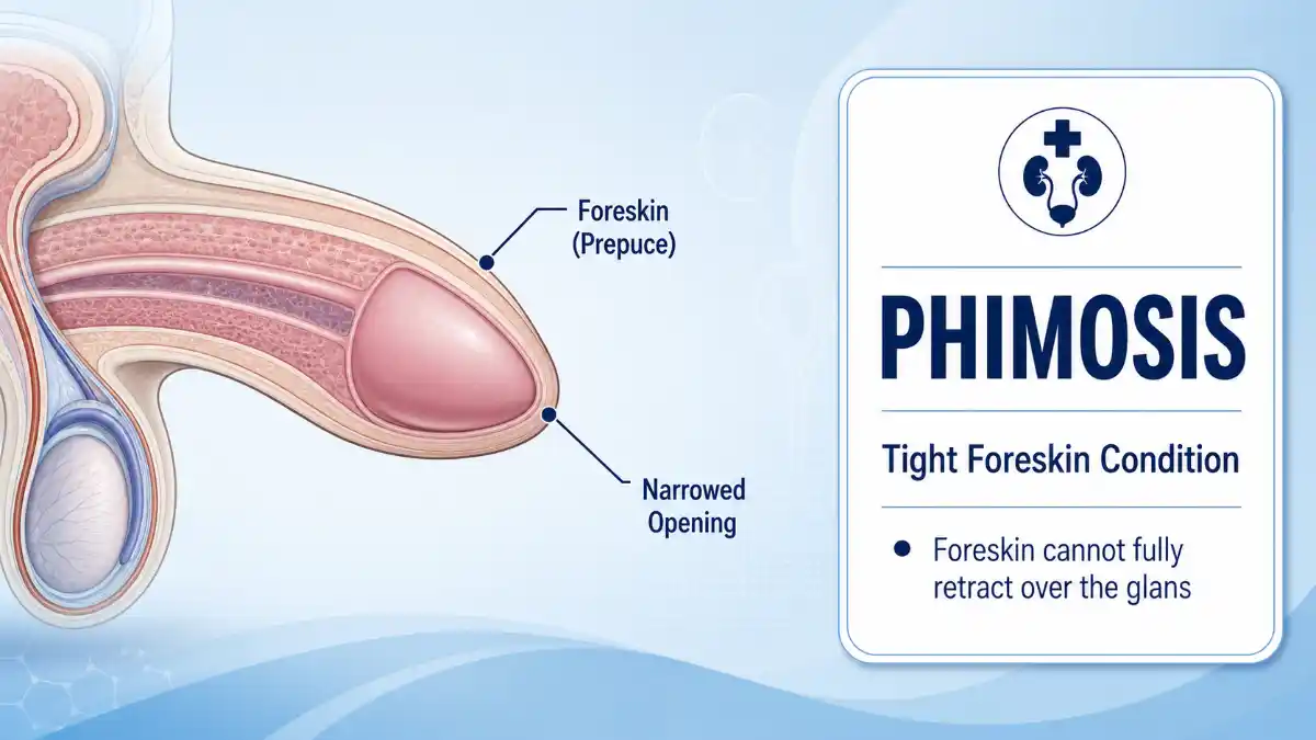
    Phimosis is a condition in which the foreskin cannot be fully pulled back over the head of the penis (glans). In babies and young boys, a non-retractable foreskin is usually a normal part of development. In older children and adults, however, new or symptomatic tightness may be related to inflammation, injury, or scarring and may need medical assessment.

    The key issue is not simply whether the foreskin retracts. Pain, bleeding, recurrent inflammation, difficulty urinating, painful erections, scarring, or a change from previously normal retraction are more important signs that evaluation may be needed.

    What Is Phimosis?

    Phimosis means that the foreskin, or prepuce, cannot be retracted sufficiently to expose the glans. A tight foreskin is not automatically a disease. Many children have a naturally non-retractable foreskin for several years, and some do not achieve full retraction until later childhood or adolescence.

    The distinction between normal developmental tightness and pathologic disease is important:

    • Physiologic phimosis is normal developmental tightness, especially in babies and young children.
    • Pathologic phimosis develops when inflammation, repeated injury, or scarring makes the foreskin abnormally tight.

    A person may therefore have a non-retractable foreskin without needing treatment. Treatment becomes more relevant when tightness causes symptoms or is associated with scarring or recurrent inflammation.

    The Two Main Types of Phimosis

    Understanding the type helps determine whether observation, medication, or a procedure is appropriate.

    Physiologic Phimosis

    Physiologic phimosis is common in babies and young children. At birth, the foreskin is normally attached to the glans and cannot usually be pulled back completely. Separation and increased retractability occur gradually as a child grows.

    A child with no pain, infection, bleeding, or difficulty urinating usually does not need treatment simply because the foreskin does not retract.

    Parents should never forcibly retract a child’s foreskin. Forced retraction can injure the tissue and contribute to inflammation or scarring.

    Pathologic Phimosis

    Pathologic phimosis occurs when the foreskin becomes abnormally tight because of inflammation, repeated irritation, trauma, or scar tissue.

    Possible warning signs include:

    • A white or hardened scar-like ring at the foreskin opening
    • Recurrent cracking or bleeding
    • Increasing tightness after previously normal retraction
    • Repeated balanitis or balanoposthitis
    • Painful erections
    • Difficulty passing urine

    In adults, lichen sclerosus, also known as balanitis xerotica obliterans (BXO) when it affects the penis, is an important cause of acquired scarring.

    Phimosis Symptoms

    Difficulty retracting the foreskin is the defining feature, but not everyone with a tight foreskin has symptoms that require treatment.

    Symptoms that deserve medical attention include:

    • Pain or tenderness of the foreskin or glans
    • Redness or swelling
    • Pain or difficulty while urinating
    • A weak or altered urine stream
    • Recurrent urinary tract infections
    • Bleeding or cracks in the foreskin
    • Thick discharge or unpleasant odor
    • Recurrent inflammation
    • Painful erections
    • Pain during sexual activity
    • Difficulty maintaining hygiene because of symptomatic tightness

    Ballooning of the foreskin during urination can occur in children with a tight foreskin and is not automatically dangerous. It becomes more concerning when it occurs with pain, difficult urination, poor flow, or recurrent infections.

    Symptoms such as redness, discharge,e or irritation can also result from balanitis, inflammation of the glans, or balanoposthitis, inflammation involving both the glans and foreskin. The underlying cause should therefore be assessed rather than assuming every penile symptom is caused by phimosis.

    Phimosis in Children

    A non-retractable foreskin is usually normal in babies and young boys. There is no single age at which every child’s foreskin must become retractable. Some boys develop full retractability earlier, while others take longer.

    For young children, normal care is simple:

    • Do not forcibly pull the foreskin back.
    • Wash the outside gently with water.
    • Avoid harsh or fragranced products that irritate sensitive skin.
    • Allow the child to retract the foreskin naturally when it becomes comfortable to do so.
    • If the foreskin is retracted, it should be returned to its normal forward position.

    Medical evaluation is appropriate if a child develops pain, significant swelling, recurrent inflammation, bleeding, difficulty urinating, recurrent infections,s or obvious scarring.

    A pediatrician or pediatric urologist can determine whether the tightness is still physiologic or has become pathologic.

    Read More: Phimosis

    Phimosis in Adults

    New or worsening tightness in adulthood deserves medical evaluation, particularly when the foreskin previously retracted normally.

    Possible contributors include:

    • Recurrent balanitis or balanoposthitis
    • Lichen sclerosus
    • Other inflammatory skin conditions
    • Repeated trauma or tearing
    • Scarring
    • Recurrent infections
    • Diabetes-associated genital inflammation

    Diabetes does not mean that every adult with a tight foreskin has diabetes. However, recurrent balanitis or fungal infections can sometimes be associated with diabetes, so a clinician may consider testing when the overall history suggests it.

    Lichen sclerosus deserves particular attention because it can produce white or pale skin changes, fissures and progressive tightening. Significant scarring may require specialist treatment.

    What Causes Phimosis?

    The likely cause depends on age, symptoms, and whether the foreskin was previously retractable.

    SituationPossible explanation
    Baby or young child without symptomsNormal developmental tightness
    Child with recurrent inflammationIrritation, infection, or developing scarring
    Adult with newly acquired tightnessInflammation, trauma, skin disease, or scarring
    Tightness after repeated forceful retractionTears followed by scar formation
    White, thickened or scarred foreskinLichen sclerosus or another scarring disorder
    Recurrent balanitisInfection, irritation or an underlying condition such as diabetes

    Read More: Phimosis

    Other skin conditions, including eczema, psoriasis and lichen planus, may also affect genital skin. Sexually transmitted infections can cause genital inflammation as well, depending on the symptoms and exposure history.

    How Is Phimosis Diagnosed?

    Diagnosis is usually made from the medical history and physical examination. Laboratory tests are not automatically required.

    A clinician may ask:

    • When did the tightness begin?
    • Was the foreskin previously retractable?
    • Is there pain during erections or urination?
    • Are there recurrent infections?
    • Has the foreskin cracked or bled?
    • Are there white, hardened,d or scarred areas?
    • Is there discharge or persistent inflammation?
    • Are there symptoms that could indicate another skin condition?

    The examination may identify a tight foreskin opening, scar tissue, inflammation, lichen sclerosus, or another anatomical issue such as a short frenulum.

    Additional tests may be appropriate when symptoms suggest infection, diabetes, or another underlying condition. The exact testing depends on the individual’s history and examination.

    Phimosis Treatment Options

    Treatment depends on age, symptoms, severity,y and the underlying cause. A child with painless physiologic tightness may need reassurance rather than treatment, while significant scarring in an adult may require specialist management.

    Observation and Gentle Care

    Observation is often appropriate for children with normal developmental tightness and no troublesome symptoms.

    Gentle hygiene is important, but aggressive cleaning is unnecessary. Perfumed soaps, shower gels and other irritants may aggravate inflamed genital skin.

    For adults with mild tightness, avoiding irritation may reduce inflammation, but hygiene alone will not reliably reverse established scar tissue.

    Topical Steroid Treatment

    A clinician may prescribe a topical corticosteroid to improve the elasticity of a tight foreskin. In children, topical corticosteroids are commonly used with gentle retraction or stretching instructions and can be effective for many cases.

    Treatment schedules and steroid strengths vary, so the medication should be used according to the clinician’s instructions.

    Steroid therapy may be less successful when substantial scar tissue is present. In such cases, the underlying cause, particularly a scarring disorder, needs to be addressed.

    Do not apply a prescription-strength steroid to genital skin without appropriate medical advice.

    Gentle Foreskin Stretching

    Stretching may form part of treatment when recommended by a clinician. It should be gentle and should never involve forceful pulling.

    The goal is gradual improvement in flexibility, not creating pain or tearing. Forceful stretching can cause small injuries and potentially contribute to further scarring.

    If stretching causes bleeding, significant pain, or tearing, stop and seek medical advice.

    Treating Infection or Inflammation

    If balanitis, balanoposthitis, or another infection is present, treatment should target the specific cause.

    Depending on the diagnosis, treatment may include an antifungal medicine, antibiotics, or another appropriate therapy. Antibiotics are not a general treatment for phimosis and should not be used unless a bacterial infection is suspected or diagnosed.

    Persistent inflammation may also require assessment for an underlying skin disorder or other contributor.

    Preputioplasty

    Preputioplasty is a foreskin-preserving surgical procedure designed to widen a narrowed foreskin opening.

    It can be considered in selected patients, particularly when preserving the foreskin is desirable. The suitability of the procedure depends on the location and severity of narrowing and whether significant scarring or an active skin condition is present.

    Circumcision

    Circumcision removes the foreskin and can provide definitive treatment for persistent symptomatic or scarred disease.

    It may be considered when other treatments have failed, when significant scarring is present, or when conditions such as lichen sclerosus have affected the foreskin. It is not automatically necessary for every person who cannot retract the foreskin.

    The decision should consider the cause of the tightness, symptoms, treatment response, anatomy, and the patient’s preferences.

    Phimosis vs. Paraphimosis

    Phimosis and paraphimosis are different conditions.

    Phimosis: The foreskin cannot be pulled back sufficiently over the glans.

    Paraphimosis: The foreskin has been pulled behind the glans but becomes trapped there and cannot be returned to its normal position.

    Paraphimosis can cause progressive swelling and restrict blood flow to the glans. It is a medical emergency and requires urgent treatment.

    If the foreskin is stuck behind the glans and cannot be moved forward again, seek emergency medical care rather than trying repeated forceful manipulation at home.

    Possible Complications

    Many cases of childhood physiologic tightness cause no lasting problems. Complications are more concerning when the foreskin is symptomatic, scarred, or repeatedly inflamed.

    Potential complications include:

    • Recurrent balanitis or balanoposthitis
    • Painful erections
    • Pain during sex
    • Recurrent urinary infections
    • Difficulty urinating
    • Progressive scarring
    • Paraphimosis
    • Urinary obstruction in severe cases

    Lichen sclerosus is particularly important because chronic genital disease can cause progressive scarring and requires appropriate medical management. Persistent abnormal skin changes, ulcers, unexplained bleeding, lumps,s or other concerning lesions should be assessed promptly rather than attributed automatically to a tight foreskin.

    When Should You See a Urologist?

    A routine medical appointment is appropriate when:

    • Tightness develops for the first time in adulthood.
    • The foreskin becomes progressively tighter.
    • Retraction causes significant pain.
    • Erections or sexual activity become painful.
    • Recurrent balanitis occurs.
    • The foreskin repeatedly cracks or bleeds.
    • The opening appears white, hardened, or scarred.
    • Urination becomes painful or difficult.
    • Conservative treatment has not worked.

    A general practitioner or primary-care clinician can often begin the assessment and refer to urology when specialist treatment is appropriate.

    When Is Phimosis an Emergency?

    Seek urgent medical care if:

    • The foreskin is stuck behind the glans and cannot be returned forward.
    • You cannot pass urine.
    • The penis becomes severely swollen or discoloured.
    • There is severe pain associated with a trapped foreskin.
    • Significant infection symptoms occur with rapidly worsening swelling or systemic illness.

    A trapped retracted foreskin may indicate paraphimosis, which can compromise blood flow and requires prompt treatment.

    Frequently Asked Questions

    Is phimosis normal in babies?

    Yes. A non-retractable foreskin is usually normal in babies and young children because the foreskin naturally separates from the glans over time. It does not require treatment simply because it cannot yet be pulled back.

    At what age should a child’s foreskin retract?

    There is no universal deadline. Many boys can retract the foreskin by around age 5, but some do not achieve full retraction until age 10 or later. Symptoms, scarring, and urinary problems are more important than using a rigid age cutoff.

    Can adults develop phimosis suddenly?

    Yes. Adult-onset tightness can occur after inflammation, recurrent infections, trauma, or scarring disorders such as lichen sclerosus. New tightness after previously normal retraction should be assessed rather than treated as normal childhood-type phimosis.

    Is ballooning of the foreskin during urination dangerous?

    Not necessarily. Ballooning can occur in children with a naturally tight foreskin. It deserves assessment when it is accompanied by pain, difficult urination, poor urinary flow, recurrent infections,s or other symptoms.

    Can phimosis go away without surgery?

    Often, yes. Painless physiologic tightness in children may resolve naturally. When treatment is necessary, prescribed topical corticosteroids and appropriate gentle retraction may work in many children. Surgery is reserved for selected cases.

    Are phimosis exercises safe?

    Gentle stretching may be used as part of clinician-directed treatment, but forceful retraction is unsafe. Stretching that causes tearing, bleeding, ng or significant pain can worsen scarring and should be stopped.

    Can phimosis affect sex?

    Yes. In adults, a tight or scarred foreskin can cause painful erections, discomfort during intercourse, or tearing of the foreskin. These symptoms are reasons to seek medical assessment because several treatment options may be available.

    Is circumcision the only treatment?

    No. Depending on the cause and severity, management may include observation, treatment of inflammation or infection, prescribed topical corticosteroids, selected foreskin-preserving procedures, or circumcision.

    Can a tight foreskin cause urinary problems?

    Yes. Significant narrowing can cause painful or difficult urination and, in severe cases, urinary obstruction. Difficulty passing urine should be medically assessed rather than managed through forceful retraction.

    Is a foreskin stuck behind the glans the same as phimosis?

    No. A foreskin that cannot be pulled back is phimosis. A foreskin that has been pulled back and becomes trapped behind the glans is paraphimosis. Paraphimosis can restrict blood flow and requires urgent medical treatment.

  • VASA Fitness Guide: Membership Cost, Locations, Hours & Amenities

    VASA Fitness Guide: Membership Cost, Locations, Hours & Amenities

    VASA Fitness is a large U.S. gym chain with 70+ locations and three core membership levels: Basic, Fitness, and STUDIO. Current advertised starting prices commonly begin at $14.99 per month for Basic, $24.99 for Fitness, and $44.99 for STUDIO, although enrollment charges, promotions, taxes,s and available amenities can vary by club.

    The best VASA Fitness membership depends on how you train. Basic is built for one-club cardio and strength workouts. Fitness adds access to all VASA locations, premium amenities, and eligible Group Fitness classes. STUDIO is for members who want structured, coach-led strength, HIIT, and infrared-yoga sessions.

    VASA Fitness locations

    VASA Fitness operates locations across 11 U.S. states, including Arizona, Colorado, Idaho, Illinois, Indiana, Iowa, Michigan, Nebraska, Oklahoma, Utah,h and Wisconsin. Some additional clubs are listed as coming soon, so confirm that a specific location is open before visiting or joining.

    The network has a strong presence in Colorado and Utah, alongside locations in expanding markets across the Midwest and Southwest. Because individual clubs differ in size, facilities, and schedules, the location page for your chosen gym should be the final reference point.

    How to find a VASA Fitness near you

    Use the official location finder and open the individual club page before enrolling. It should confirm:

    • Club address and phone number
    • Whether the location is open, pre-sale,e or coming soon
    • Current VASA Fitness hours
    • Membership prices and local promotions
    • Group Fitness and STUDIO schedules
    • Pool, sauna, court, and recovery-area availability
    • KidCare availability and operating hours

    Amenities are location-specific. A club with a pool, sauna, basketball court, or infrared-yoga studio may offer a very different experience from another VASA Fitness location.

    VASA Fitness membership cost

    VASA Fitness offers three main membership options. The monthly price is important, but it is not the full cost of joining. Enrollment fees, the Rate Guarantee Fee, taxes,s and optional services can all affect the total amount you pay.

    MembershipCurrent advertised starting price*Main accessBest for
    Basic$14.99/monthOne home club and core cardio/strength equipmentIndependent training at one convenient gym
    Fitness$24.99/monthAll 70+ VASA Fitness locations, premium amenities and eligible Group Fitness classesMembers who want flexibility and broader facility access
    STUDIO$44.99/monthFitness benefits plus eligible boutique-style STUDIO classesPeople who use coached classes regularly

    Read More: VASA Fitness

    *Membership rates, enrollment charges, promotions,s and amenities may differ by location.

    Which VASA plan is best?

    Choose the lowest-priced plan that best fits how you will actually use the gym.

    • Basic: Best for one-club members focused on weights, machines, and cardio.
    • Fitness: Best for members who want all-club access, Group Fitness, s,s and premium amenities.
    • STUDIO: Best for people who regularly attend coached strength, HIIT, or infrared-yoga sessions.

    A higher-tier membership is not automatically the better value. If you rarely use classes, pools, courts,s or recovery spaces, a more expensive plan may not improve your workout experience enough to justify the added monthly cost.

    What does VASA cost upfront?

    The advertised monthly rate is only the starting point. Your actual cost when joining can include an enrollment or down-payment charge, the annual Rate Guarantee Fee, applicable taxes, and optional services.

    First-year cost = (monthly membership × 12) + enrollment fee + Rate Guarantee Fee + applicable taxes + optional services

    For example, a $24.99 monthly Fitness membership equals $299.88 in base dues over 12 months. That does not include enrollment charges, annual fees, taxes, KidCare, personal training, or other optional purchases.

    Enrollment charges can vary by club and membership tier. Some locations run introductory offers that reduce or waive certain upfront charges, while others may use a higher Basic enrollment amount. Always review the final checkout screen for your selected club.

    Rate Guarantee Fee

    VASA’s published terms state that members who joined on or after September 1, 2025 pay a $59.99 Rate Guarantee Fee per member, plus applicable sales tax. The fee is billed two months after joining and then annually while the membership remains active.

    Before submitting payment, review the membership agreement carefully and save a copy of the dues, fees, notice requirements, and billing schedule.

    What Basic membership includes

    Basic is VASA Fitness’s entry-level option. It is designed for members who want independent training at one designated home club without paying for premium amenities or classes.

    Basic members can typically use:

    • One home VASA Fitness location
    • Cardio equipment and standard workout areas
    • Free weights
    • Strength-training machines
    • Functional-training turf
    • Performance-lifting areas where available

    Basic is a practical choice if the club is close to your home or workplace and your workouts are focused on lifting, cardio, and self-directed training.

    What Basic does not include

    Basic membership is intentionally limited. It does not include premium facilities and services such as:

    • Pools, hot tubs, saunas, spas, or steam rooms
    • Group Fitness classes
    • Basketball or racquetball courts
    • Massage lounges
    • Tanning or red light therapy
    • STUDIO LFT, STUDIO RED, or STUDIO FLOW

    Basic members are also restricted to one home club. A home-club transfer can carry a $25 fee, so choose your primary location carefully before joining.

    What Fitness membership includes

    Fitness is VASA’s middle-tier plan. It is aimed at members who want more flexibility and more facility access than Basic provides.

    Fitness includes access to all 70+ VASA Fitness locations, premium amenities, and eligible Group Fitness classes. The exact equipment, amenities, and schedule can still vary by club, so multi-location access does not mean every gym has the same pool, court, sauna, or group class timetable.

    Depending on the location, Fitness members may have access to:

    • Indoor pools, hot tubs, saunas and steam rooms
    • Basketball and racquetball courts
    • Group Fitness classes
    • HydroMassage lounges, tanning and selected recovery amenities
    • KidCare as an optional service
    • Guest privileges under current plan and location terms

    Fitness is often the most balanced choice for people who train at more than one club, enjoy group workouts, or want access to aquatic and recovery amenities.

    What STUDIO membership includes

    STUDIO is VASA Fitness’s premium membership tier. It includes Fitness benefits, along with unlimited access to eligible boutique-style STUDIO classes at locations where they are available.

    The main STUDIO formats include:

    • STUDIO LFT: Coach-led strength training
    • STUDIO RED: High-intensity interval training
    • STUDIO FLOW: Infrared yoga

    STUDIO may also include additional benefits such as InBody scans and eligible KidCare discounts. These are useful extras, but the primary reason to choose STUDIO is regular access to coached workouts.

    STUDIO booking rules

    STUDIO availability varies by location. Before upgrading, check whether your preferred club offers the format and timetable you want.

    Cancel STUDIO reservations at least 12 hours before class to avoid a late-cancellation charge. The first no-show in a month costs $3, while subsequent no-shows that month cost $10 each.

    VASA Fitness hours

    VASA Fitness does not have one universal operating schedule. Each club sets its own hours, which can change for holidays, maintenance, or other operational reasons.

    Some locations offer early-morning through late-night access on weekdays, with shorter weekend hours. However, those times should never be treated as a network-wide schedule.

    Separate operating hours may also apply to:

    • KidCare
    • Group Fitness classes
    • STUDIO sessions
    • Pools and hot tubs
    • Saunas, steam rooms and recovery spaces
    • Personal-training appointments

    Check the individual club page before visiting, especially if you train early, late, or during holidays.

    VASA Fitness amenities

    VASA Fitness amenities should be compared by location and membership tier. Instead of assuming every club offers every feature, focus on the facilities you will use most.

    CategoryTypical amenitiesWhat to verify
    TrainingCardio decks, free weights, machines, turf and lifting areasEquipment selection and crowding at your workout time
    Aquatics and recoveryPools, hot tubs, sauna, steam room and massage loungesWhether the amenity exists and is included in your tier
    SportsBasketball and racquetball courtsCourt availability, rules and access requirements
    ServicesKidCare, personal training and InBody scansAdd-on costs, reservations and operating hours
    ClassesGroup Fitness and STUDIO programmingMembership eligibility and local schedule

    Read More: VASA Fitness

    Basic is primarily for core workout equipment. Fitness adds access to a broader range of amenities and eligible Group Fitness. STUDIO adds eligible boutique-style coaching.

    Is VASA Fitness worth it?

    VASA Fitness can be a strong value option for people who want a larger gym, relatively low advertised monthly pricing, and the option to access premium amenities or classes.

    Basic is most worthwhile for one-club cardio and strength training. Fitness is usually better for members who need access to multiple clubs, Group Fitness, pools, courts,s or recovery areas. STUDIO is most useful for people who value accountability and will attend coach-led sessions regularly.

    Before joining, visit at the time you expect to work out. Look at parking, rush-hour crowding, locker-room condition, equipment availability, ty and whether the class schedule fits your week. A low monthly rate only matters if the gym works with your real routine.

    Membership policies to know

    Cancellation

    VASA memberships must be cancelled in accordance with the process and notice requirements set out in the individual membership agreement. Current terms generally require 30 days’ notice. A $25 cancellation-processing fee can apply if a standard month-to-month membership is cancelled within the first six months, subject to applicable exceptions and state-specific rules.

    Membership freeze

    VASA allows members to freeze a membership for up to 90 days. A $15 fee applies per freeze request. During the freeze, gym access and billing pause, and the membership automatically reactivates after the freeze period ends.

    Downgrading a plan

    A $25 downgrade fee can apply when moving from Fitness or STUDIO to a lower membership tier within the first six months. Consider this before upgrading solely for a short-term promotion or a class format you may not use consistently.

    Free 1-day pass

    VASA currently promotes a free 1-day gym pass. Eligibility and club-specific conditions may apply, so check the current offer and identification requirements at your chosen location before visiting.

    Frequently asked questions

    How many VASA Fitness locations are there?

    VASA Fitness has 70+ locations across 11 U.S. states, with additional clubs listed as coming soon. The directory may include active, presale, and future facilities, so check that your nearest location is open before planning a visit or buying a membership.

    How much is VASA Fitness per month?

    VASA Fitness memberships commonly start at $14.99 per month for Basic, $24.99 for Fitness, and $44.99 for STUDIO. Those are starting rates. Enrollment charges, annual fees, taxes, promotions, and optional services can change the final cost.

    What does Basic membership not include?

    Basic does not include pools, saunas, steam rooms, Group Fitness, basketball, racquetball, massage lounges, tanning, red light therapy,y or STUDIO programming. It is designed for one-club access to core strength and cardio equipment.

    Does Fitness membership include all VASA locations?

    Yes. Fitness provides access to all 70+ VASA Fitness locations. Amenities, class schedules,s and services can vary by club, so check your preferred locations if you need a particular pool, court, sauna, or fitness class.

    Are STUDIO classes available at every VASA Fitness?

    No. STUDIO members receive access to eligible classes, but the available formats and schedules differ by location. Confirm that your club offers STUDIO LFT, STUDIO RE, D, or STUDIO FLOW before upgrading.

    What happens if I miss a STUDIO class?

    Cancel at least 12 hours before the class to avoid a late-cancellation charge. The first no-show in a month costs $3, while subsequent no-shows in the same month cost $10 each.

    Can I freeze a VASA Fitness membership?

    Yes. VASA allows memberships to be frozen for up to 90 days. Each freeze request has a $15 fee, and gym access and billing pause while the membership is frozen.

    Can I change my VASA Fitness home club?

    Basic members can request a home-club transfer through VASA support. A $25 transfer fee can apply. If you regularly train at more than one gym, Fitness may be a better fit because it includes all-club access.

    Does VASA Fitness charge an annual fee?

    Yes. The Rate Guarantee Fee is $59.99 per member plus applicable sales tax for members who joined on or after September 1, 2025. It is billed two months after joining and annually thereafter.

    Is VASA Fitness worth joining?

    VASA Fitness is worth considering if its location, hours,s and facilities match your lifestyle. Basic suits independent training at one gym; Fitness is better for flexibility and broader amenities; and STUDIO is ideal for members who use coached classes frequently.

  • Xiphoid Process: Anatomy, Location, Function, Pain & Symptoms

    Xiphoid Process: Anatomy, Location, Function, Pain & Symptoms

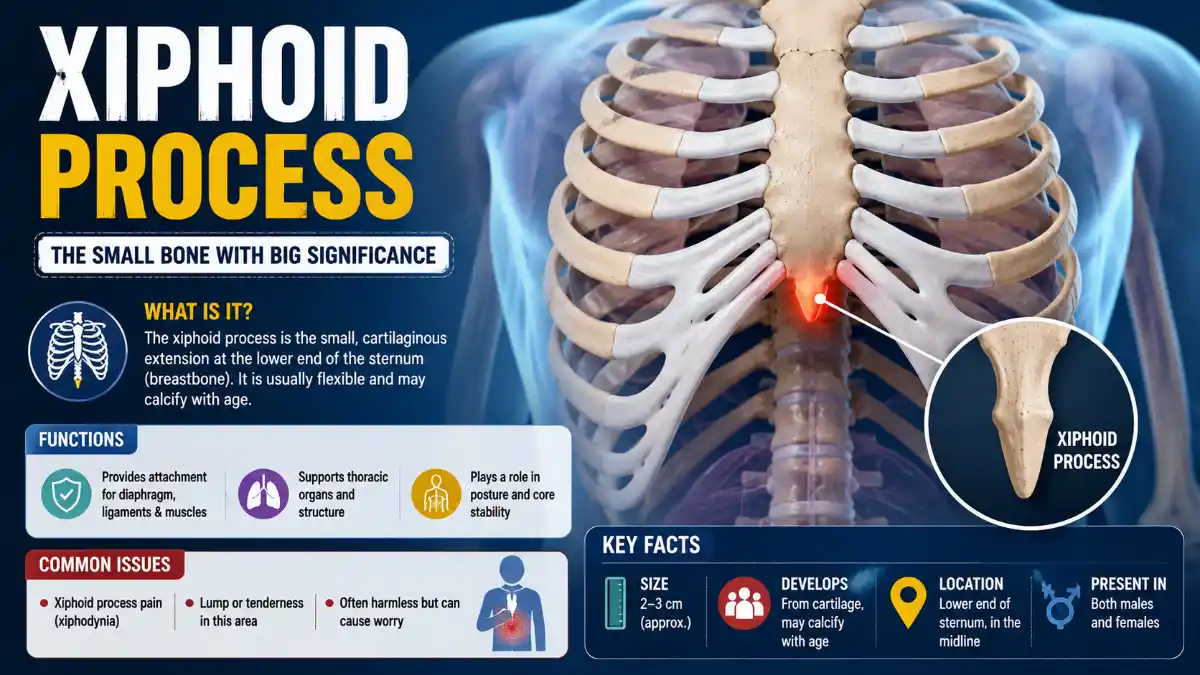
    The xiphoid process is the small, pointed segment at the very bottom of the sternum (breastbone). It sits in the soft area where the lower ribs meet at the center of the chest. It anchors several abdominal and chest wall muscles and also serves as an important positioning landmark during CPR.

    The xiphoid process can become tender due to strain, trauma, or a condition called xiphodynia. However, problems such as acid reflux or gallbladder disease can also cause pain in the same region without involving the xiphoid process itself. This guide explains its anatomy, location, clinical significance, and how true xiphoid pain differs from other pain felt in this area.

    What Is the Xiphoid Process?

    The xiphoid process, also called the xiphisternum or ensiform process, is the smallest and most inferior of the three sections that make up the sternum, alongside the manubrium and the sternal body. It is purely cartilaginous at birth, and ossification generally begins in the superior portion around age three. Ossification timing and the maturity of the xiphisternal joint vary considerably between individuals – some people show a fully bony joint by roughly age 40, while others retain some cartilage well beyond that.

    FeatureDetail
    Part ofSternum (third and most inferior segment)
    ShapeTypically triangular; commonly varies – can be broad, thin, curved, bifid, trifid, or contain small foramina
    LengthApproximately 2–5 cm in adults
    Tissue at birthCartilage
    OssificationBegins around age 3 in most people, though timing varies widely between individuals
    Approximate vertebral levelLevel of the T10 vertebra

    Read More: Xiphoid

    Because shape and ossification vary so widely, an unusually curved, forked, or prominent tip seen on imaging is often a normal anatomical variant rather than a mass. Studies using CT imaging have found that a majority of people have some degree of deflection or a non-straight shape, which is why isolated variation rarely signals a problem on its own.

    Where Is the Xiphoid Process Located?

    It sits at the lowest tip of the sternum, in the epigastric region of the chest, at approximately the level of the T10 vertebra. You can locate it by running a finger down the center of the breastbone until it ends in a small, pointed or notch-like projection, just above where the ribcage curves inward toward the abdomen.

    It connects to the sternal body above through the xiphisternal joint, has facets for part of the seventh costal cartilage, and attaches to the linea alba, the fibrous midline seam running down the front of the abdominal wall, at its inferior tip.

    What Does the Xiphoid Process Do?

    Its main role is structural: this bone anchors several muscles and ligaments rather than performing an active function on its own. Structures attached to it include:

    • Rectus abdominis – fibers of this abdominal muscle attach at its anterior surface
    • Internal and external oblique aponeuroses – connective tissue sheets from the abdominal wall
    • Diaphragmatic slips – part of the muscle responsible for breathing, attached posteriorly
    • Transversus thoracis – a deep chest wall muscle
    • Costoxiphoid ligaments – formed with the seventh costal cartilage, stabilizing the lower ribcage

    Because of these attachments, this small structure indirectly supports breathing mechanics and core stability, even though it doesn’t move independently.

    The Xiphoid Process in CPR

    It’s an anatomical landmark to avoid during chest compressions; correct hand placement is the center of the chest, over the lower half of the sternum, above the tip rather than on it. Current American Heart Association guidance for adult CPR directs rescuers to compress the center of the chest on the lower half of the sternum. It’s important not to press directly on the tip itself, since this bone is comparatively soft and can fracture under compression, potentially injuring the liver, heart, or diaphragm. For complete, up-to-date instructions on hand placement and compression technique, refer to current CPR training resources rather than a general anatomy guide.

    What Is Xiphodynia (Xiphoid Syndrome)?

    Xiphodynia, also called xiphoidalgia or xiphoid syndrome, is an uncommon condition in which an irritated xiphoid process or xiphisternal joint produces tenderness and pain that can radiate to the chest, upper abdomen, throat, or arms. Because this referred pain pattern overlaps with several more common and more dangerous conditions, xiphodynia is frequently under-recognized or misdiagnosed.

    Typical presentation includes localized tenderness at the base of the sternum that may worsen with pressing on the area, bending, twisting, deep breathing, or lying face-down. Diagnosis is primarily clinical. During a physical examination, reproduction of the patient’s typical pain when a clinician palpates the tender area can support the diagnosis, but this finding is not conclusive on its own and shouldn’t be treated as a self-test.

    Differential diagnosis matters greatly, since this syndrome’s symptom pattern can be mistaken for – or coexist with – angina, myocardial infarction, pericarditis, pulmonary embolism, peptic ulcer disease, or gallbladder disease. Clinicians are trained to complete a careful history and physical exam, and to exclude cardiac and other serious causes, before attributing symptoms to this area.

    Management is typically conservative, and there is no standardized treatment protocol given how uncommon and under-studied the condition is. Approaches described in the clinical literature include activity modification and clinician-directed conservative care; some case reports describe local anesthetic or steroid injections improving symptoms. Xiphoidectomy, the surgical removal of the affected bone, is reserved for rare, confirmed, refractory cases after conservative measures fail.

    Xiphoid-Area Pain: Origin vs. Overlapping Causes

    Pain felt near the lower sternum can come from this structure itself, or from a nearby organ whose pain simply overlaps with the region – and telling the two apart usually requires clinical evaluation, not self-diagnosis.

    Causes that can originate at or directly involve this area:

    • Mechanical strain from heavy lifting, intense core exercise, or persistent coughing that stresses the muscle and ligament attachments
    • Direct trauma from a fall, sports injury, or steering-wheel impact, which can bruise or, less commonly, fracture the bone
    • Xiphodynia, as described above
    • Costochondritis affecting cartilage near the lower sternum, which can produce pain radiating toward this region

    Conditions that can mimic or overlap with pain here, without being true xiphoid pathology:

    • Gastroesophageal reflux disease (GERD) and peptic ulcer disease, which sit anatomically close to this area and are frequently considered – and ruled out – before a xiphodynia diagnosis is made
    • Gallbladder disease (cholelithiasis)
    • Cardiac conditions, including angina and myocardial infarction
    • Pulmonary embolism

    Xiphoid-Area Pain vs. GERD vs. Cardiac Pain

    FeatureChest-wall originGERDPossible cardiac pain
    Reproduced by clinician pressing on the areaOftenUsually notUsually not
    Changes with torso movement or breathingOftenLess typicalVariable
    Worse after meals or lying downLess typicalCommonVariable
    Burning sensationPossibleCommonPossible
    Shortness of breath, sweating, or radiating arm/jaw painNot typicalNot typicalWarning sign
    Requires emergency assessmentUsually noUsually noPotentially yes

    This table is a general guide, not a diagnostic tool. Overlapping or atypical presentations are common, and any new or severe chest or upper-abdominal pain should be medically evaluated rather than matched against a checklist.

    How Xiphoid-Area Pain Is Evaluated

    A clinician typically starts with a history of when the pain began, what triggers or relieves it, and any recent injury. This is followed by a physical examination, including gentle palpation to check whether pressing on the tender spot reproduces the pain. Depending on the findings, the clinician may evaluate for cardiac, gastrointestinal, or pulmonary causes through additional questions, examination, or targeted testing. Imaging, such as an X-ray, ultrasound, or CT scan, is generally reserved for cases involving significant trauma, a palpable mass, suspected fracture, or an unclear diagnosis after the initial workup.

    Swollen Xiphoid Process or Lump: What It Usually Means

    A noticeable lump or firmness at the bottom of the sternum is most often this bone becoming more prominent rather than a new growth. Contributing factors include weight loss, which reduces the soft tissue covering the area; pregnancy, as the growing uterus pushes against the lower ribcage; natural variation in how far the tip curves or flares; and localized swelling following an injury.

    A new lump, rapidly growing, hard, fixed in place, or accompanied by skin changes is not typical of a simple prominent tip and warrants clinical evaluation to determine the cause.

    When to Seek Medical Evaluation

    Seek emergency care immediately for sudden or severe chest pain accompanied by shortness of breath, sweating, dizziness, or pain radiating to the arm or jaw, as these can indicate a cardiac event.

    Seek routine medical evaluation for pain or a lump that is new, persistent beyond a few weeks, worsening, linked to a significant chest injury, or accompanied by fever or skin changes. A clinician can typically distinguish chest-wall pain from cardiac, digestive, or gallbladder causes through history, physical examination, and imaging when indicated.

    Key Takeaways

    This small structure at the base of the sternum anchors abdominal and chest wall muscles, serves as a CPR landmark, and can occasionally become a genuine source of pain through xiphodynia, strain, or trauma. Pain felt in this region can also originate from nearby structures or conditions, such as GERD or gallbladder disease, rather than from the bone itself. Distinguishing between the two – and ruling out cardiac causes first – is a job for clinical evaluation rather than self-diagnosis.

    Frequently Asked Questions

    Can you normally feel your xiphoid process, or does it mean something is wrong?

    Yes, it’s normal to feel this structure, particularly in people with less chest fat or a naturally prominent tip. Feeling it isn’t a sign of a problem by itself. A newly appearing lump, rapid growth, or pain accompanying it are the features that warrant a medical evaluation, not the ability to palpate the bone itself.

    How do doctors diagnose xiphodynia?

    Diagnosis relies mainly on clinical examination. A clinician checks whether palpating the tender area reproduces the patient’s typical pain, which can support the diagnosis, though it isn’t conclusive on its own. Because the condition’s symptom pattern overlaps with cardiac, gastrointestinal, and other thoracic conditions, clinicians typically rule out more dangerous causes first through history, physical exam, and appropriate testing before settling on this diagnosis.

    Can acid reflux cause pain near the xiphoid process?

    GERD can cause pain felt in the same general area, since the lower esophagus and upper stomach sit nearby, but this is generally considered a condition that overlaps with or mimics chest-wall pain rather than direct injury to the bone itself. Clinicians often consider and rule out GERD when evaluating symptoms in this region before diagnosing a xiphoid-specific cause such as xiphodynia.

    Is a protruding xiphoid process dangerous?

    A protruding or curved tip is usually a normal anatomical variation rather than a dangerous finding; imaging studies have found that a majority of people have some degree of deflection or a non-straight shape. It becomes a concern mainly if it’s linked to new pain, a recent injury, or if it’s mistaken for an abnormal mass, in which case a clinician can confirm through examination or imaging that it’s a normal variant.

    Can doctors remove the xiphoid process if it causes ongoing problems?

    In rare, confirmed cases of xiphodynia that don’t respond to conservative management, a procedure called xiphoidectomy can be performed to remove the affected bone. This is uncommon and reserved for refractory cases after other treatment options and other diagnoses have been thoroughly considered.

  • Maxilla Bone Explained: Structure, Function, Fractures & Related Anatomy

    Maxilla Bone Explained: Structure, Function, Fractures & Related Anatomy

    The Facial bone is a paired bone that forms your upper jaw, and it does far more than hold your teeth in place. Sitting at the center of the midface, this bone shapes the floor of the eye sockets, the walls of the nasal cavity, and the roof of the mouth – all while anchoring the muscles used to chew, speak, and smile.

    This guide covers the Facial bone’s structure, its important anatomical landmarks, its neurovascular relationships, and its clinical significance in trauma, dentistry, and reconstructive surgery.

    What Is the Facial Bone?

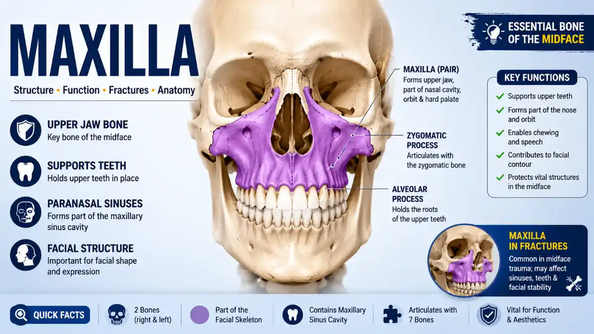
    The Facial bone is a paired, pyramidal bone that forms the upper jaw and the central portion of the facial skeleton. The two Facial bones fuse at the midline along the median maxillary bony suture, together forming what’s commonly called “the Facial bone.” It’s the second-largest bone of the face, surpassed in size only by the mandible.

    The Facial bone is fixed in position, unlike the mandible, which moves at the temporomandibular joint. This fixed structure gives the lower jaw a stable surface to work against during chewing, and it makes the Facial bone a reliable anatomical reference point during trauma assessment and reconstructive planning.

    Together, the two Facial bones contribute to four major facial structures: the hard palate, the floor and lateral wall of the nasal cavity, the floor of the orbit, and the maxillary sinus – the largest of the four paranasal sinuses.

    Facial bone Anatomy at a Glance

    FeatureDetails
    LocationMidface, forming the upper jaw
    TypePaired, pyramidal facial bone
    Number of bonesTwo (fused at the median maxillary bony suture)
    Main partsOne body, four processes
    Four surfaces of bodyAnterior, posterior (infratemporal), superior (orbital), medial (nasal)
    Four processesFrontal, zygomatic, palatine, alveolar
    Articulates withEight named neighboring bones plus the opposite Facial bone
    ContainsMaxillary bony sinus, the largest paranasal sinus
    Primary nerve supplyMaxillary branch of the trigeminal nerve (CN V2) and its branches
    Core functionsAnchors upper teeth, shapes midface, forms part of orbit and nasal cavity, supports speech and mastication

    Anatomical Structure of the Facial Bone

    Body and Four Surfaces

    The body of the Facial bone is pyramidal and hollow, housing the maxillary sinus. It has four surfaces, each carrying distinct landmarks:

    • Anterior (facial) surface – includes the infraorbital foramen, canine fossa, and incisive fossa
    • Posterior (infratemporal) surface – faces the infratemporal fossa; contains the maxillary bony tuberosity and alveolar foramina
    • Superior (orbital) surface – forms most of the orbital floor; contains the infraorbital groove and canal
    • Medial (nasal) surface – forms part of the lateral nasal wall; contains the maxillary bony ostium and conchal crest

    Four Processes

    ProcessLocationKey Articulation
    Frontal processExtends upward and mediallyArticulates with the frontal and nasal bones; forms the lacrimal groove with the lacrimal bone
    Zygomatic processExtends laterallyArticulates with the zygomatic bone; forms the superolateral border of the maxillary sinus
    Palatine processExtends mediallyForms roughly two-thirds of the hard palate and part of the nasal floor
    Alveolar processExtends downwardHorseshoe-shaped; forms the tooth sockets and extends posteriorly to the maxillary bony tuberosity

    Read More: Maxilla

    The alveolar process forms the maxillary bony dental arch – the row of eight sockets on each side that hold the upper teeth. Its bone density and shape directly affect procedures like extractions, implants, and orthodontic treatment, and it undergoes progressive resorption after tooth loss, which is why long-term edentulism changes facial profile and denture fit over time.

    Important Anatomical Landmarks of the Facial Bone

    Beyond the body and processes, several smaller landmarks are clinically significant and frequently referenced in anatomy education:

    • Infraorbital foramen, groove, and canal – the groove crosses the orbital floor, continues as the infraorbital canal, and opens at the infraorbital foramen roughly 1 cm below the orbital rim; it transmits the infraorbital nerve and vessels. Trauma here often causes cheek and upper-lip numbness.
    • Canine fossa – a depression on the anterior surface lateral to the canine eminence; serves as the origin of the levator anguli oris muscle.
    • Incisive fossa – a shallow depression medial to the canine eminence on the anterior surface.
    • Anterior nasal spine – a pointed projection formed by the palatine processes at the base of the nasal cavity.
    • Incisive canal and foramen – located in the anterior hard palate; transmits the nasopalatine nerve and sphenopalatine artery branches, connecting the oral and nasal cavities.
    • Maxillary bony tuberosity – a rounded prominence at the posterior end of the alveolar process; anchors part of the pterygoid musculature and marks a common fracture reference point.
    • Alveolar foramina – small openings on the infratemporal surface transmitting the posterior superior alveolar nerves and vessels to the molars.
    • Lacrimal groove – formed with the lacrimal bone on the frontal process; continues inferiorly as part of the nasolacrimal canal.
    • Maxillary bony ostium (Maxillary bony hiatus) – the drainage opening of the maxillary bony sinus into the nasal cavity’s middle meatus.
    • Orbital floor contribution and relation to the inferior orbital fissure – the Facial bone forms most of the orbital floor; the inferior orbital fissure runs along its posterolateral margin and separates it from the greater wing of the sphenoid, a relevant landmark in orbital floor fractures.

    What Bones Does the Facial bone Articulate With?

    Each Facial bone articulates with eight named neighboring bones plus the opposite Facial bone, giving nine total articulations when the contralateral Facial bone is included. This extensive connectivity is part of why fractures here can affect such a wide area of the face:

    • Frontal bone (superiorly)
    • Nasal bone
    • Lacrimal bone
    • Ethmoid bone
    • Zygomatic bone (laterally)
    • Inferior nasal concha
    • Vomer
    • Palatine bone (posteriorly)
    • Opposite Facial bone (at the midline)

    Neurovascular Relationships of the Facial bone

    Nerve supply. Sensation to the Facial bone comes from the facial nerve (CN V2), the second division of the trigeminal nerve. After leaving the skull through the foramen rotundum, it enters the pterygopalatine fossa and gives off several branches: the infraorbital nerve (its direct continuation, exiting at the infraorbital foramen to supply the lower eyelid, nose, cheek, and upper lip), the posterior, middle, and anterior superior alveolar nerves (supplying the upper molars, premolars, and incisors respectively, together with the sinus lining), and the greater palatine and nasopalatine nerves (supplying the hard palate).

    Blood supply. Arterial supply comes mainly from branches of the Facial bony artery, a terminal branch of the external carotid artery. Its pterygopalatine segment gives rise to the posterior superior alveolar, infraorbital, descending palatine, and sphenopalatine arteries, which together supply the Facial bony teeth, sinus lining, palate, and lateral nasal wall.

    Understanding this neurovascular network matters clinically: injury to the infraorbital nerve is a common cause of midface numbness after trauma, and awareness of the arterial arcade around the Facial bonery sinus helps surgeons minimize bleeding during sinus and dental procedures.

    The Facial Bonery Sinus

    The Facial bonery sinus, housed within the body of the Facial bone, is the largest of the four paranasal sinuses. It is pyramidal in shape, with its base forming part of the lateral nasal wall and its apex pointing toward the zygomatic process. It drains into the nasal cavity’s middle meatus through the maxillary bony ostium. Reported adult sinus volumes vary considerably between individuals, sex, and measurement method, so no single figure applies universally.

    At birth, the sinus is very small, expanding considerably through childhood and adolescence as the face grows. Proposed functions of the paranasal sinuses include reducing skull weight and contributing to the conditioning (warming and humidifying) of inhaled air and to vocal resonance, though the precise physiological significance of the sinuses is still debated among anatomists.

    Because the sinus floor sits close to the roots of the upper premolars and molars – sometimes separated by only a thin plate of bone – dental infections can spread into the sinus, and sinus infections can mimic tooth pain. This overlap is clinically important for both dentists and ENT specialists.

    Functions of the Facial Bone

    Mastication (Chewing)

    The alveolar process anchors the upper teeth, giving the mandible’s teeth a fixed surface to grind against. Chewing forces are distributed through reinforced facial buttresses – most notably the nasomaxillary buttresses and zygomaticomaxillary buttresses – toward the cranial skeleton, which is why this region is dense enough to withstand repeated mechanical load throughout life.

    Speech and Facial Expression

    The facial bones anchor several muscles of facial expression, including the nasalis, levator labii superioris, levator labii superioris alaeque nasi, and levator anguli oris – muscles involved in lip movement, nostril dilation, and smiling. The volume and shape of the hard palate also influence how sound resonates during speech.

    Breathing and Sinus Function

    By forming the floor and part of the lateral wall of the nasal cavity, the Facial bone helps direct airflow and supports structures involved in filtering and conditioning inhaled air, with the Facial bonery sinus contributing to this process as outlined above.

    Supporting the Orbit and Protecting Nerves

    The Facial bone forms most of the orbital floor and transmits branches of the Facial bonery nerve, including the infraorbital nerve. Damage to this region – from trauma or surgery – can cause numbness across the cheek, upper lip, and side of the nose.

    How the Facial Bone Develops

    Facial development begins around the fourth week of gestation, when paired facial bones and mandibular prominences form from the first pharyngeal arch, alongside a single frontonasal prominence, around the primitive mouth. The maxillary bony prominences give rise to most of the Ffacial bones, the secondary palate, and the lateral parts of the upper lip, forming mainly through intramembranous ossification with a brief secondary cartilage contribution near the zygomatic process.

    The secondary palate forms when the paired palatal shelves of the facial bones’ prominences rotate into a horizontal position and fuse at the midline, a process generally complete by the tenth to twelfth week of gestation. When this fusion fails, the result is a cleft palate, sometimes with an accompanying cleft lip.

    Common Facial Bone Injuries and Conditions

    Facial Bone Fractures

    Because the facial bones connect to so many surrounding bones, fractures here rarely stay contained to one spot – they often extend into the nose, orbit, or cheekbone. Facial bone fractures are classified using the Le Fort system, based on fracture patterns first described in 1901:

    • Le Fort I – a horizontal fracture separating the alveolar and palatine processes (with the teeth) from the rest of the Facial bone
    • Le Fort II – a pyramidal fracture running from the nasal bridge through the frontal process, infraorbital margin, and maxillary bony sinus
    • Le Fort III – craniofacial dissociation, separating the entire facial skeleton from the skull base via the zygomatic arches and orbital floors

    Symptoms typically include facial swelling, bruising around the eyes, malocclusion (difficulty aligning the bite), nosebleeds, and – in more severe fractures involving the orbital floor – vision changes or facial numbness from nerve involvement. CT imaging is the standard method for confirming fracture pattern and guiding surgical planning. Recovery timelines vary considerably with severity and treatment approach and should be assessed individually by the treating surgeon; uncomplicated fractures generally heal faster than those requiring surgical fixation, with full functional recovery – including bite alignment and any nerve-related numbness – often trailing behind bone healing itself. Any patient with facial trauma and associated vision changes should seek urgent evaluation.

    Sinus-Related Issues

    Because the maxillary sinus floor sits so close to the tooth roots, infections can travel in either direction. Odontogenic sinusitis occurs when a dental infection spreads upward into the sinus, producing symptoms that resemble ordinary sinusitis – facial pressure, nasal congestion, tenderness – but which require dental rather than purely medical treatment to resolve.

    Cleft Palate and Congenital Anomalies

    When the palatal shelves of the two maxillary bony prominences fail to fuse completely, the result is a cleft palate, sometimes with a cleft lip. These conditions can affect feeding, speech development, and occasionally hearing, and are typically managed with a combination of surgical repair, speech therapy, and long-term dental care.

    Facial bone vs. Mandible: Key Differences

    FeatureFacial boneMandible
    PositionUpper jawLower jaw
    MobilityFixedMobile, via the temporomandibular joint
    Number of bonesPaired, fused at the midlineSingle, fused bone
    SinusContains the maxillary bony sinusNo sinus
    FormationIntramembranous ossificationPrimarily intramembranous ossification adjacent to Meckel’s cartilage, with secondary cartilage contributing to specific regions such as the condyle
    Fracture patternOften multi-bone (Le Fort fractures)Typically localized to the angle, body, or condyle

    Why the Facial Bone Matters in Dentistry and Surgery

    The Facial bone’s bone density and its proximity to the sinus directly influence treatment planning. Dental implants placed in the upper posterior teeth sometimes require a sinus-lift procedure when the maxillary sinus floor sits too close to the available bone, a common consequence of long-term tooth loss. The specific amount of bone height needed before a sinus lift is recommended varies by implant system, grafting technique, and individual anatomy, so this decision is made case by case by an oral surgeon or periodontist rather than following a single fixed rule.

    In orthodontics, the width and growth pattern of the upper jaw affect bite alignment and palate shape. Palatal expansion is generally more predictable in younger patients before advanced skeletal maturation of the midpalatal suture; treatment options for skeletally mature patients – such as surgically assisted expansion – may differ. Understanding the Facial bone’s relationship to the temporomandibular joint and surrounding musculature also guides orthognathic surgery planning for patients with significant bite discrepancies.

    Key Takeaways

    • The Facial bone is a paired, pyramidal bone forming the upper jaw and the structural core of the midface.
    • It consists of a body with four surfaces and four processes (frontal, zygomatic, palatine, alveolar), articulating with eight named bones plus the opposite Facial bone.
    • It houses the maxillary sinus and transmits branches of the maxillary nerve (CN V2), including the infraorbital nerve.
    • Its extensive articulations mean fractures often extend across multiple facial bones, classified using the Le Fort system.
    • Its close relationship with the maxillary sinus creates a two-way link between dental and sinus health.
    • Clinical procedures such as sinus lifts and palatal expansion depend directly on its anatomy and growth pattern, with treatment decisions made on an individual basis.

    Frequently Asked Questions

    What is the difference between the Maxillary bone and the Maxillary bonery sinus?

    The Maxillary bone is the bone itself; the Maxillary bonery sinus is the air-filled cavity within its body. As the largest paranasal sinus, it drains into the nasal cavity through the Maxillary bonery ostium. It is very small at birth and enlarges through childhood, which is why sinus-related dental and ENT issues become more common with age rather than in early childhood.

    Can you feel your maxillary bone?

    Yes. It forms the bony ridge beneath your cheeks and around your nose, plus the hard portion of the palate just behind your upper front teeth. The alveolar process holding your upper teeth is also palpable through the gum tissue.

    How long does it take for a maxillary bony fracture to heal?

    It depends on severity and treatment approach, and should be assessed individually by the treating surgeon. Simple, non-surgical fractures generally heal faster than complex Le Fort fractures requiring surgical fixation, and full functional recovery – including bite alignment and resolution of any nerve-related numbness – often takes longer than the underlying bone healing itself.

    Does the Maxillary bone grow after childhood?

    It grows most actively during childhood and adolescence, largely finishing by the late teens to early twenties as the midpalatal suture and other growth sites mature. This is why interventions like palatal expansion tend to work best before advanced skeletal maturity.

    What nerve provides sensation to the Maxillary bone?

    The maxillary nerve (CN V2), the second division of the trigeminal nerve, supplies the upper jaw, teeth, and midface. Its infraorbital branch is especially relevant clinically, since it is commonly affected in orbital floor fractures and certain dental procedures, and injury can cause cheek, lip, and nasal numbness.

    Why does a maxillary sinus infection sometimes feel like a toothache?

    The floor of the maxillary sinus sits very close to – sometimes just a thin bony plate away from – the roots of the upper molars and premolars. Sinus inflammation can therefore produce pain that feels dental in origin, which is why clinicians sometimes rule out sinus involvement before treating upper-back-tooth pain with no obvious cause.

    What causes a cleft palate in the Maxillary bone?

    A cleft palate results when the palatal shelves of the two maxillary bones fail to fuse at the midline during fetal development, a process normally completed by around the tenth to twelfth week of pregnancy. The cause is usually multifactorial, involving genetic predisposition alongside environmental factors such as maternal nutrition or certain medication exposures.

    Is the Maxillary bone one bone or two?

    It consists of two separate maxillary bones fused at the median maxillary suture, though they are commonly referred to together as “the Maxillary bone” in clinical and everyday language.

    What is a sinus lift, and why is it related to the Maxillary bone?

    A sinus lift is a surgical procedure that adds bone to the posterior Maxillary bone by elevating the floor of the Maxillary bone’s sinus, typically performed before placing dental implants in patients with reduced bone height in this region. The need for the procedure and the amount of bone added depend on individual anatomy and implant planning rather than a single universal measurement.

    Can a fractured Maxillary bone affect vision?

    Yes, particularly in fractures involving the orbital floor that the Maxillary bone helps form. Vision changes can result from swelling, nerve injury, or entrapment of the eye muscles at the fracture site. Facial trauma accompanied by vision changes, double vision, or restricted eye movement requires urgent medical evaluation.