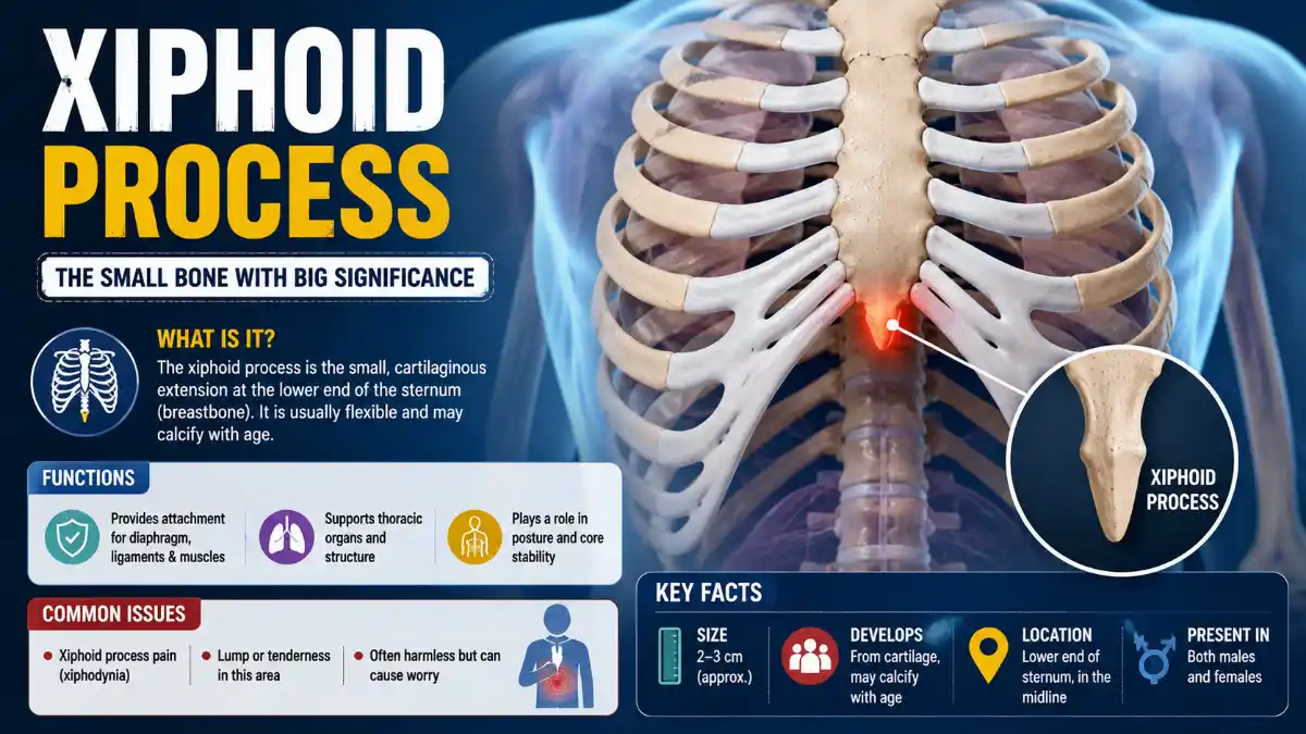

The xiphoid process is the small, pointed segment at the very bottom of the sternum (breastbone). It sits in the soft area where the lower ribs meet at the center of the chest. It anchors several abdominal and chest wall muscles and also serves as an important positioning landmark during CPR.
The xiphoid process can become tender due to strain, trauma, or a condition called xiphodynia. However, problems such as acid reflux or gallbladder disease can also cause pain in the same region without involving the xiphoid process itself. This guide explains its anatomy, location, clinical significance, and how true xiphoid pain differs from other pain felt in this area.
What Is the Xiphoid Process?
The xiphoid process, also called the xiphisternum or ensiform process, is the smallest and most inferior of the three sections that make up the sternum, alongside the manubrium and the sternal body. It is purely cartilaginous at birth, and ossification generally begins in the superior portion around age three. Ossification timing and the maturity of the xiphisternal joint vary considerably between individuals – some people show a fully bony joint by roughly age 40, while others retain some cartilage well beyond that.
| Feature | Detail |
| Part of | Sternum (third and most inferior segment) |
| Shape | Typically triangular; commonly varies – can be broad, thin, curved, bifid, trifid, or contain small foramina |
| Length | Approximately 2–5 cm in adults |
| Tissue at birth | Cartilage |
| Ossification | Begins around age 3 in most people, though timing varies widely between individuals |
| Approximate vertebral level | Level of the T10 vertebra |
Read More: Xiphoid
Because shape and ossification vary so widely, an unusually curved, forked, or prominent tip seen on imaging is often a normal anatomical variant rather than a mass. Studies using CT imaging have found that a majority of people have some degree of deflection or a non-straight shape, which is why isolated variation rarely signals a problem on its own.
Where Is the Xiphoid Process Located?
It sits at the lowest tip of the sternum, in the epigastric region of the chest, at approximately the level of the T10 vertebra. You can locate it by running a finger down the center of the breastbone until it ends in a small, pointed or notch-like projection, just above where the ribcage curves inward toward the abdomen.
It connects to the sternal body above through the xiphisternal joint, has facets for part of the seventh costal cartilage, and attaches to the linea alba, the fibrous midline seam running down the front of the abdominal wall, at its inferior tip.
What Does the Xiphoid Process Do?
Its main role is structural: this bone anchors several muscles and ligaments rather than performing an active function on its own. Structures attached to it include:
- Rectus abdominis – fibers of this abdominal muscle attach at its anterior surface
- Internal and external oblique aponeuroses – connective tissue sheets from the abdominal wall
- Diaphragmatic slips – part of the muscle responsible for breathing, attached posteriorly
- Transversus thoracis – a deep chest wall muscle
- Costoxiphoid ligaments – formed with the seventh costal cartilage, stabilizing the lower ribcage
Because of these attachments, this small structure indirectly supports breathing mechanics and core stability, even though it doesn’t move independently.
The Xiphoid Process in CPR
It’s an anatomical landmark to avoid during chest compressions; correct hand placement is the center of the chest, over the lower half of the sternum, above the tip rather than on it. Current American Heart Association guidance for adult CPR directs rescuers to compress the center of the chest on the lower half of the sternum. It’s important not to press directly on the tip itself, since this bone is comparatively soft and can fracture under compression, potentially injuring the liver, heart, or diaphragm. For complete, up-to-date instructions on hand placement and compression technique, refer to current CPR training resources rather than a general anatomy guide.
What Is Xiphodynia (Xiphoid Syndrome)?
Xiphodynia, also called xiphoidalgia or xiphoid syndrome, is an uncommon condition in which an irritated xiphoid process or xiphisternal joint produces tenderness and pain that can radiate to the chest, upper abdomen, throat, or arms. Because this referred pain pattern overlaps with several more common and more dangerous conditions, xiphodynia is frequently under-recognized or misdiagnosed.
Typical presentation includes localized tenderness at the base of the sternum that may worsen with pressing on the area, bending, twisting, deep breathing, or lying face-down. Diagnosis is primarily clinical. During a physical examination, reproduction of the patient’s typical pain when a clinician palpates the tender area can support the diagnosis, but this finding is not conclusive on its own and shouldn’t be treated as a self-test.
Differential diagnosis matters greatly, since this syndrome’s symptom pattern can be mistaken for – or coexist with – angina, myocardial infarction, pericarditis, pulmonary embolism, peptic ulcer disease, or gallbladder disease. Clinicians are trained to complete a careful history and physical exam, and to exclude cardiac and other serious causes, before attributing symptoms to this area.
Management is typically conservative, and there is no standardized treatment protocol given how uncommon and under-studied the condition is. Approaches described in the clinical literature include activity modification and clinician-directed conservative care; some case reports describe local anesthetic or steroid injections improving symptoms. Xiphoidectomy, the surgical removal of the affected bone, is reserved for rare, confirmed, refractory cases after conservative measures fail.
Xiphoid-Area Pain: Origin vs. Overlapping Causes
Pain felt near the lower sternum can come from this structure itself, or from a nearby organ whose pain simply overlaps with the region – and telling the two apart usually requires clinical evaluation, not self-diagnosis.
Causes that can originate at or directly involve this area:
- Mechanical strain from heavy lifting, intense core exercise, or persistent coughing that stresses the muscle and ligament attachments
- Direct trauma from a fall, sports injury, or steering-wheel impact, which can bruise or, less commonly, fracture the bone
- Xiphodynia, as described above
- Costochondritis affecting cartilage near the lower sternum, which can produce pain radiating toward this region
Conditions that can mimic or overlap with pain here, without being true xiphoid pathology:
- Gastroesophageal reflux disease (GERD) and peptic ulcer disease, which sit anatomically close to this area and are frequently considered – and ruled out – before a xiphodynia diagnosis is made
- Gallbladder disease (cholelithiasis)
- Cardiac conditions, including angina and myocardial infarction
- Pulmonary embolism
Xiphoid-Area Pain vs. GERD vs. Cardiac Pain
| Feature | Chest-wall origin | GERD | Possible cardiac pain |
| Reproduced by clinician pressing on the area | Often | Usually not | Usually not |
| Changes with torso movement or breathing | Often | Less typical | Variable |
| Worse after meals or lying down | Less typical | Common | Variable |
| Burning sensation | Possible | Common | Possible |
| Shortness of breath, sweating, or radiating arm/jaw pain | Not typical | Not typical | Warning sign |
| Requires emergency assessment | Usually no | Usually no | Potentially yes |
This table is a general guide, not a diagnostic tool. Overlapping or atypical presentations are common, and any new or severe chest or upper-abdominal pain should be medically evaluated rather than matched against a checklist.
How Xiphoid-Area Pain Is Evaluated
A clinician typically starts with a history of when the pain began, what triggers or relieves it, and any recent injury. This is followed by a physical examination, including gentle palpation to check whether pressing on the tender spot reproduces the pain. Depending on the findings, the clinician may evaluate for cardiac, gastrointestinal, or pulmonary causes through additional questions, examination, or targeted testing. Imaging, such as an X-ray, ultrasound, or CT scan, is generally reserved for cases involving significant trauma, a palpable mass, suspected fracture, or an unclear diagnosis after the initial workup.
Swollen Xiphoid Process or Lump: What It Usually Means
A noticeable lump or firmness at the bottom of the sternum is most often this bone becoming more prominent rather than a new growth. Contributing factors include weight loss, which reduces the soft tissue covering the area; pregnancy, as the growing uterus pushes against the lower ribcage; natural variation in how far the tip curves or flares; and localized swelling following an injury.
A new lump, rapidly growing, hard, fixed in place, or accompanied by skin changes is not typical of a simple prominent tip and warrants clinical evaluation to determine the cause.
When to Seek Medical Evaluation
Seek emergency care immediately for sudden or severe chest pain accompanied by shortness of breath, sweating, dizziness, or pain radiating to the arm or jaw, as these can indicate a cardiac event.
Seek routine medical evaluation for pain or a lump that is new, persistent beyond a few weeks, worsening, linked to a significant chest injury, or accompanied by fever or skin changes. A clinician can typically distinguish chest-wall pain from cardiac, digestive, or gallbladder causes through history, physical examination, and imaging when indicated.
Key Takeaways
This small structure at the base of the sternum anchors abdominal and chest wall muscles, serves as a CPR landmark, and can occasionally become a genuine source of pain through xiphodynia, strain, or trauma. Pain felt in this region can also originate from nearby structures or conditions, such as GERD or gallbladder disease, rather than from the bone itself. Distinguishing between the two – and ruling out cardiac causes first – is a job for clinical evaluation rather than self-diagnosis.
Frequently Asked Questions
Can you normally feel your xiphoid process, or does it mean something is wrong?
Yes, it’s normal to feel this structure, particularly in people with less chest fat or a naturally prominent tip. Feeling it isn’t a sign of a problem by itself. A newly appearing lump, rapid growth, or pain accompanying it are the features that warrant a medical evaluation, not the ability to palpate the bone itself.
How do doctors diagnose xiphodynia?
Diagnosis relies mainly on clinical examination. A clinician checks whether palpating the tender area reproduces the patient’s typical pain, which can support the diagnosis, though it isn’t conclusive on its own. Because the condition’s symptom pattern overlaps with cardiac, gastrointestinal, and other thoracic conditions, clinicians typically rule out more dangerous causes first through history, physical exam, and appropriate testing before settling on this diagnosis.
Can acid reflux cause pain near the xiphoid process?
GERD can cause pain felt in the same general area, since the lower esophagus and upper stomach sit nearby, but this is generally considered a condition that overlaps with or mimics chest-wall pain rather than direct injury to the bone itself. Clinicians often consider and rule out GERD when evaluating symptoms in this region before diagnosing a xiphoid-specific cause such as xiphodynia.
Is a protruding xiphoid process dangerous?
A protruding or curved tip is usually a normal anatomical variation rather than a dangerous finding; imaging studies have found that a majority of people have some degree of deflection or a non-straight shape. It becomes a concern mainly if it’s linked to new pain, a recent injury, or if it’s mistaken for an abnormal mass, in which case a clinician can confirm through examination or imaging that it’s a normal variant.
Can doctors remove the xiphoid process if it causes ongoing problems?
In rare, confirmed cases of xiphodynia that don’t respond to conservative management, a procedure called xiphoidectomy can be performed to remove the affected bone. This is uncommon and reserved for refractory cases after other treatment options and other diagnoses have been thoroughly considered.


[…] the proximal phalanx. The PIP and DIP joints occur farther along the finger. The thumb has only one interphalangeal joint because it lacks a middle […]