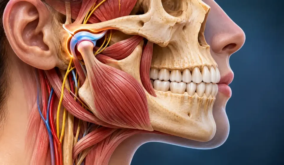

Jaw anatomy includes the upper jaw (maxilla), lower jaw (mandible), temporomandibular joints (TMJs), muscles, nerves, blood vessels, and tooth-supporting structures. The maxilla forms the stationary upper jaw, while the mandible is the movable lower jaw. Together, these structures support chewing, speaking, swallowing, facial shape, and dental alignment.
The mandible connects to the skull through two temporomandibular joints located just in front of the ears. Muscles control movement at these joints, while the teeth provide surfaces for biting and chewing. Nerves carry sensory information and control the chewing muscles, and blood vessels supply the bones and surrounding tissues. Muscles beneath the jaw also play an important role in swallowing.
Major Structures of the Human Jaw
Several structures work together to make normal jaw movement, sensation, and dental function possible.
| Anatomical Structure | Location | Primary Function |
| Maxilla | Upper jaw | Supports upper teeth and forms part of the facial skeleton |
| Mandible | Lower jaw | Supports lower teeth and enables jaw movement |
| Temporomandibular joints | In front of each ear | Connect the mandible to the skull |
| Jaw muscles | Around the cheeks, temples, and lower jaw | Control chewing and jaw movement |
| Jaw nerves | Throughout the jaws and surrounding tissues | Provide sensation and motor control |
| Blood vessels | Within and around the jaw | Supply oxygen and nutrients |
| Alveolar bone | Around tooth roots | Forms sockets supporting the teeth |
| Periodontal ligament | Between tooth roots and their sockets | Anchors teeth and cushions biting forces |
The bones provide the jaw’s framework, but movement depends on the muscles and joints. Teeth, nerves, and supporting tissues contribute to biting, chewing, and other everyday oral functions.
Jaw Bone Anatomy: Mandible and Maxilla
The mandible and maxilla form the bony foundation of the jaws. Both support teeth, but they differ in structure, mobility, and how they connect to the skull.
Mandible Anatomy: The Lower Jaw
The mandible is the largest and strongest facial bone. Its curved horizontal body forms the chin and lower dental arch, while two vertical sections, called rami, extend upward toward the temporomandibular joints. The anatomy of the mandible includes several landmarks that help explain its movement, muscle attachments, and nerve pathways.
Important mandibular landmarks include:
- Body: The horizontal portion that supports the lower teeth.
- Ramus: The vertical portion that provides attachment sites for chewing muscles.
- Angle: The junction where the body meets the ramus.
- Condylar process: The posterior projection that forms the mandibular part of the TMJ.
- Coronoid process: The anterior projection where the temporalis muscle attaches.
- Mandibular notch: The curved depression between the condylar and coronoid processes.
- Mandibular symphysis: The midline region where the two halves of the developing mandible fuse.
- Mental protuberance: The bony prominence that forms the chin.
- Alveolar portion: The tooth-bearing bone containing the lower tooth sockets.
- Mandibular foramen: An opening on the inner surface of the ramus where the inferior alveolar nerve and blood vessels enter.
- Mandibular canal: The passage within the bone that carries these nerves and blood vessels.
- Mental foramen: An opening on the outer surface of the mandibular body where the mental nerve and accompanying blood vessels emerge.
These landmarks are especially important in dentistry because they help clinicians locate nerves, assess fractures, and understand how the jaw moves.
Maxilla Anatomy: The Upper Jaw
The maxilla consists of two bones fused at the midline. Together, they support the upper teeth and form much of the central facial skeleton.
Its main anatomical features include:
- Body: Contains the maxillary sinus, an air-filled cavity within the upper jaw.
- Alveolar process: Holds the sockets for the upper teeth.
- Palatine process: Forms the anterior portion of the hard palate, or roof of the mouth.
- Frontal process: Extends upward toward the frontal bone.
- Zygomatic process: Connects with the cheekbone.
The maxilla also helps form the nasal cavity and the floor of each eye socket. Unlike the mandible, it does not move independently during chewing.
Mandible vs. Maxilla: Key Differences
| Feature | Mandible | Maxilla |
| Position | Lower jaw | Upper jaw |
| Mobility | Movable | Stationary |
| Structure | Single adult bone | Two fused bones |
| Connection | Articulates with temporal bones through TMJs | Joined to surrounding facial bones by sutures |
| Teeth supported | Lower teeth | Upper teeth |
| Main function | Jaw movement and biting | Upper dental support and facial structure |
Temporomandibular Joint (TMJ) Anatomy
The temporomandibular joint connects the mandibular condyle to the temporal bone on each side of the skull, just in front of the ear. Unlike a simple hinge, the TMJ combines rotation with sliding movements, allowing the lower jaw to move in several directions.
Each TMJ contains several important structures:
- Mandibular condyle: The rounded upper end of the mandible that participates in the joint.
- Mandibular fossa: A depression in the temporal bone that accommodates the condyle.
- Articular eminence: A bony prominence that helps guide the condyle during forward movement.
- Articular disc: A fibrocartilaginous structure that separates the joint into upper and lower compartments.
- Joint capsule: Connective tissue that encloses the joint and helps maintain its stability.
- Supporting ligaments: Structures that reinforce the joint and limit excessive movement.
The TMJ produces two main types of movement: rotation and translation. Rotation occurs primarily in the lower joint compartment, while translation allows the condyle and disc to slide forward along the temporal bone.
Both movements occur together during normal jaw function. The temporomandibular joint’s anatomy and movement explain how people can open their mouths, chew, speak, and move the lower jaw from side to side.
Jaw Muscles and Their Functions
Four primary muscles of mastication control most chewing movements. These muscles attach to the skull and mandible and receive motor signals from the mandibular division of the trigeminal nerve.
The main muscles of mastication have different actions, although they work together during everyday movements.
| Muscle | Location or Attachment | Primary Action |
| Masseter | Cheekbone to mandibular ramus and angle | Closes the jaw |
| Temporalis | Temple region to coronoid process | Elevates and retracts the mandible |
| Medial pterygoid | Deep facial region to inner mandibular ramus | Elevation, protrusion, and sideways movement |
| Lateral pterygoid | Deep facial region to the pterygoid fovea on the mandibular neck, with variable attachments to the TMJ disc and capsule | Protrusion, translation, and assisted jaw opening |
Other muscles assist with opening the mouth and swallowing.
The digastric helps lower the mandible when the hyoid bone is stabilized. The mylohyoid supports the floor of the mouth and helps elevate the hyoid, while the geniohyoid moves the hyoid forward and assists in lowering the jaw.
These muscles must work in coordination. For example, closing the mouth involves several muscles contracting together, while opening it requires a different combination of muscle activity and joint movement.
Jaw Nerves and Blood Supply
The jaw contains an extensive network of nerves responsible for sensation and muscle control. Much of this nerve supply comes from the trigeminal nerve, also called cranial nerve V.
Two divisions are particularly important:
Maxillary nerve (V2): Provides sensation to the upper teeth, upper gums, and surrounding facial structures.
Mandibular nerve (V3): Carries sensory information from the lower jaw and motor signals to the primary chewing muscles.
Several smaller branches supply specific areas of the mouth and face. The mandibular nerve and its branches are particularly important in understanding lower jaw sensation and movement.
Important branches include:
- Inferior alveolar nerve: Travels through the mandibular canal and supplies the lower teeth.
- Mental nerve: Supplies sensation to the chin, lower lip, and nearby tissues.
- Lingual nerve: Provides general sensation to the anterior two-thirds of the tongue and parts of the mouth.
- Buccal nerve: Supplies sensation to the cheek lining and gums near the lower molars.
- Superior alveolar nerves: Supply the upper teeth and associated dental tissues.
- Auriculotemporal nerve: Carries sensory information from the TMJ and nearby temporal region.
The inferior alveolar nerve is especially important in dental care. Because it travels inside the mandible, dentists must consider its location when administering local anesthesia, removing impacted wisdom teeth, or performing certain jaw procedures.
Blood Supply of the Jaw
The maxillary artery supplies many deep facial and jaw structures. One of its branches, the inferior alveolar artery, enters the mandibular foramen and supplies the lower jaw and teeth.
The facial artery provides blood to superficial facial tissues and areas around the mouth. Branches of the superficial temporal and maxillary arteries also contribute to the TMJ’s blood supply.
Together, these vessels deliver oxygen and nutrients to the bones, muscles, and surrounding tissues, supporting normal function and tissue repair.
Teeth, Jaw Alignment, and Supporting Structures
The upper and lower jaws contain alveolar processes, which form the bony sockets surrounding tooth roots. Each tooth is connected to its surrounding alveolar bone by the periodontal ligament, a specialized connective tissue that helps hold the tooth in place and distribute biting forces.
Dental occlusion describes how the upper and lower teeth contact each other when the jaws close.
Biting and chewing depend on more than tooth position alone. The relationship between the dental arches, jaw alignment, chewing muscles, and TMJs determines how the teeth come together during movement.
Although teeth are anchored within the jawbones, they have their own anatomical structures. Enamel covers the crown, dentin forms much of the tooth, and the pulp contains nerves and blood vessels. These tissues are distinct from the surrounding jawbone.
Why Jaw Anatomy Matters Clinically
Understanding the jaw’s structure also helps explain common dental problems, joint conditions, and injuries.
Temporomandibular disorders (TMD) can affect the jaw joints, chewing muscles, and surrounding tissues. Clicking sometimes occurs when the articular disc moves differently relative to the mandibular condyle. However, painless clicking is common and does not necessarily indicate a disorder.
According to guidance on temporomandibular disorders, clicking without pain generally does not require treatment. Painful clicking, jaw locking, or difficulty opening the mouth should be evaluated by a healthcare professional.
Mandibular fractures can involve the body, angle, or condylar region, sometimes damaging nearby nerves and blood vessels. A jaw dislocation occurs when the mandibular condyle moves out of its normal position and cannot return normally.
Malocclusion refers to an abnormal relationship between the upper and lower teeth. Impacted wisdom teeth may also lie close to the inferior alveolar nerve, making their position important when planning dental procedures.
Clinicians choose imaging methods based on the structures they need to examine. Dental X-rays and panoramic radiographs show teeth and surrounding bone, while CT or cone-beam CT provides more detailed views of bony anatomy. MRI can help evaluate the TMJ disc and other soft tissues when medically indicated.
Read also: Jaw Anatomy
Frequently Asked Questions
Can Losing Teeth Change the Shape of the Jawbone?
Yes. After a tooth is lost, the surrounding alveolar bone may gradually shrink because it no longer receives the same forces through the tooth and periodontal ligament. This process, called bone resorption, can change the shape and height of the jaw ridge over time. The amount of bone loss varies between individuals.
Why Can the Lower Lip Feel Numb After Dental Work?
Numbness may occur because local anesthetic temporarily blocks signals in the inferior alveolar or mental nerve. Sensation usually returns as the anesthetic wears off. Less commonly, irritation or injury to a nearby nerve during a dental procedure can cause longer-lasting numbness. If sensation does not return as expected or numbness persists, contact your dentist or oral surgeon.


[…] Head and neck muscles allow for chewing, making facial expressions, and positioning your head. The masseter is responsible for closing your jaw, while the sternocleidomastoid assists with turning and bending your neck. Your knowledge of where these two muscles are located relative to each other and surrounding tissue will increase as you study jaw anatomy. […]