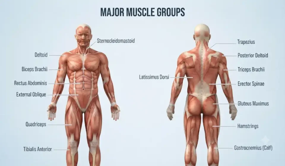

A muscle diagram will display all of the different muscles within the human body. Typically, a muscle diagram will contain two separate views of the body: an anterior view and a posterior view. This makes identifying the larger skeletal muscles easier, for example, the deltoids, pectorals, biceps, abdominals, quadriceps, glutes, hamstrings, and calves. All of these muscles assist in maintaining posture and producing movement by working together with bones, joints,s and tendons.
The body of a person includes over 600 skeletal muscles; however, the exact number will depend upon how each is classified. Generally, most of the body’s major muscle groups are shown on full-body charts, but not all of the individual muscle structures. Learning where each muscle is found and its function can assist in reading and studying anatomical charts. Additionally, understanding the way in which the muscular system is anatomically arranged may help to explain why muscles are named as such.
Labeled Human Muscle Diagram: Front and Back Views
The muscle diagram of the body with both an anterior (front) view and a posterior (back) view displays the major superficial muscles that are closest to the skin of the body. An anterior view will show the major muscles of the chest, abdomen, arms, ms and the front portion of each leg. A posterior view will show the major muscles of the back, shoulders, buttocks, and the rear portions of each leg.
Anterior (Front) Muscle Diagram
Anterior views illustrate some of the most common daily functions of muscles. Examples include: flexing the elbow joint, extending the shoulder joint, and extending the knee joint.
Major muscles visible from the front include:
- The Sternocleidomastoid runs on either side of the neck to allow the head to rotate and flex.
- Deltoid: Covers the shoulder and helps lift and rotate the arm.
- Pectoralis major: Spans the upper chest and helps bring the arm across the body.
- Biceps Brachii: The muscle that is located on the anterior side of the upper arm, allows for flexion of the elbow joint, and rotation of the forearm to allow the palm to face superiorly.
- The Rectus Abdominis muscle runs vertically on the anterior surface of the abdominal wall. This muscle assists in flexing the torso forward.
- External Oblique Muscles: These are located on both sides of your abdomen; they assist in rotating and bending your torso.
- Quadriceps – Four muscles located on the top and side of the leg. These muscles are used to extend (straighten) your legs.
- The front of the shin bone has a muscle called Tibialis Anterior, which helps to raise your foot up towards your lower leg.
Posterior (Back) Muscle Diagram
The muscles identified in the posterior view function to assist with proper posture, pull the upper extremity posteriorly, extend the lower extremities, and provide control of movement at the ankle.
Important posterior muscles include:
- Trapezius: It covers most of the upper back and neck, assisting with movement and stabilization of the shoulder blades.
- Posterior Deltoid: This is the posterior part of the shoulder that allows for backward movement of the arm.
- The triceps brachii is located on the back of the upper arm and is responsible for extending the elbow.
- The latissimus dorsi is a large muscle located in the lower and middle back. It assists with pulling the arm down and back.
- Erector Spinae: A collection of muscles located on both sides of your spine that help to make your back straight and provide stability to your back.
- Gluteus maximus: A large buttock muscle that provides powerful hip extension.
- The Hamstring Muscles are a group of muscles located on the posterior aspect of the lower limb. This muscle group has two main functions: flexing the knee joint and extending the hip joint. There is one exception to this rule, which is the short head of the Biceps Femoris.
- The calf muscle (gastrocnemius) is what lifts up the heel and also bends the knee.
- The soleus is located primarily under the gastrocnemius, with some parts of it exposed to view on either side of the gastrocnemius. The soleus aids in lifting the heel off the ground, as well as providing support to keep you stable when you are either standing or walking.
Major Muscles of the Human Body and Their Functions
When you group the most important muscles in your body based on their location on your body, it is much easier to remember which muscles do what. Each area of your body has groups of muscles that work with each other to allow you to move around, control your joints, and keep your body in balance.
| Body Region | Major Muscles | Main Functions |
| Head and neck | Masseter, sternocleidomastoid | Chewing and head movement |
| Shoulders | Deltoid, rotator cuff muscles | Arm movement and shoulder stabilization |
| Chest | Pectoralis major and minor | Arm and shoulder blade movement |
| Upper arms | Biceps brachii, brachialis, triceps brachii | Elbow flexion and extension |
| Forearms | Brachioradialis, wrist flexors and extensors | Elbow and wrist movement, grip assistance |
| Abdomen | Rectus abdominis, obliques, transverse abdominis | Trunk movement and stability |
| Back | Trapezius, latissimus dorsi, rhomboids, erector spinae | Shoulder blade, arm, and spinal movement |
| Hips | Gluteus maximus, gluteus medius, iliopsoas | Hip movement and pelvic stability |
| Thighs | Quadriceps, hamstrings, adductors | Knee and hip movement |
| Lower legs | Gastrocnemius, soleus, tibialis anterior | Ankle movement and walking |
Head, Neck, Shoulders, Chest, and Arms
Head and neck muscles allow for chewing, making facial expressions, and positioning your head. The masseter is responsible for closing your jaw, while the sternocleidomastoid assists with turning and bending your neck. Your knowledge of where these two muscles are located relative to each other and surrounding tissue will increase as you study jaw anatomy.
The deltoid is responsible for movement of the upper arm, while four other rotator cuff muscles provide support to the shoulder joint and aid in its rotation. On either side of the chest, the pectoralis major brings the arm forward and across the body. These movements will be detailed through the study of the shoulder and upper-limb muscle anatomy.
The biceps brachii and brachialis are located in the upper arm, and they each contribute to the ability of the arm to move towards the shoulder (i.e., flex) at the elbow joint. The triceps brachii is also found in the upper arm; however, unlike the other two muscles, it is responsible for moving the arm away from the shoulder (i.e., extending) at the elbow joint. There are numerous muscles within the forearm that allow individuals to control movement of their wrists and fingers. These muscles provide the mechanical support required for individuals to grasp items with their hands, to write using a pen or pencil, and to complete other types of complex tasks requiring manual dexterity.
Abdomen, Back, and Hips
The abdominal muscles provide support for the torso, allowing for proper function of the spine. The rectus abdominis muscle allows for movement in front-to-back directions (flexion), whereas the internal and external oblique muscles allow for rotational movement as well as lateral movement (side bending). In addition to providing support, the transverse abdominis muscle provides structural integrity to the abdominal wall. Knowledge of the muscles of the abdominal wall assists in distinguishing between the various layers.
On your back, you have several different groups of muscles that work together to move and stabilize your upper body. Some examples include: the trapezius and rhomboid groups, which are responsible for stabilizing the scapulae (shoulder blades); the latissimus dorsi muscle, which can be used to move your arms forward or backward; and the erector spinae muscles, which provide support for extending the spine and maintaining good posture.
The hip area is composed of a number of very large, strong muscles that each perform various functions. One such muscle is the gluteus maximus, which acts to extend the hip. Another muscle, the gluteus medius, works to help maintain pelvic stability as we walk. Additionally, there is a muscle called the iliopsoas, which can be found in a deeper location in the hip area and performs the role of lifting the thigh upward towards the torso.
Thighs and Lower Legs
The quadriceps are composed of four separate muscles: the rectus femoris, the vastus lateralis, the vastus medialis, and the vastus intermedius. These four muscles work together to allow you to extend your knee joint. Extending your knee joint allows you to stand from a sitting position, walk up stairs, and generally move about on foot.
The three muscles that comprise the hamstrings are the biceps femoris, semitendinosus, and semimembranosus. Each of these muscles is involved with flexion of the knee joint. Additionally, each of them contributes to extension of the hip joint. The adductor muscles of the thigh move the legs toward the center line of the body. These two sets of muscles are crucial in the development of the knee joint, as well as maintaining stability within the joint itself.
The gastrocnemius and soleus (lower leg) both act to push the foot down toward the ground, as you would do when you rise on your toes. Conversely, the tibialis anterior does the reverse, acting to pull the front of the foot up toward your body as you walk. Additional detail concerning the interaction between the pelvic girdle and lower-limb muscles will provide more insight into the way in which they coordinate movement.
Superficial vs. Deep Muscle Diagrams
A diagram of the human muscular system can display either superficial muscles or deep muscles, or both in different anatomical planes.
Superficial muscles are located near the surface of the body and create the appearance of muscles. Some examples of superficial muscles include the deltoid, pectoralis major, rectus abdominis, and gastrocnemius.
Many of the most important muscles involved in movement and stabilization of joints are located deeper than other muscles and/or tissues. It is difficult to observe these deep muscles with a traditional full-body image; however, many of them have very significant roles in supporting and allowing motion as well as providing support to joints.
Important deep muscles include:
- Rotator cuff muscles: The supraspinatus, infraspinatus, teres minor, and subscapularis allow for movement and stability of the shoulder.
- The transverse abdominis is a deep abdominal muscle that supports both the compression of your abdomen and the stability of your trunk.
- Multifidus: These are numerous tiny muscles located on each side of the spinal column that aid in stabilizing the spine.
- Iliopsoas: A major hip flexor formed by the iliacus and psoas major.
- Deep Hip Rotator Muscles: The deep hip rotator muscles (such as the Piriformis and Obturator Internus) assist in rotating and stabilizing the hip.
A separate regional diagram can help us locate deeper structures in our body, as many of the larger superficial muscles can obscure these structures from view.
Muscle tissue can be classified into three main groups: skeletal, smooth, and cardiac. Most full-body muscle diagrams show skeletal muscles, which are used to control movement of the body’s skeleton. Smooth muscle can be seen in structures like the digestive system and blood vessels. Cardiac muscle is what forms the heart. All three of these types are included under the larger classification of muscle tissue.
How to Read and Study a Muscle Diagram
Start by identifying if you’re viewing the anterior (front) or posterior (back) aspect of the image. It is important to remember that the left and right sides of the image will correspond with the subject’s left and right sides, not yours.
Next, work with those areas you are most comfortable with, such as your shoulder, chest, stomach, back, hip,p and leg. Before trying to recall what each muscle does, first locate each one.
In addition to helping us identify each muscle individually rather than as part of a larger group of muscles, it can assist us in identifying which muscles make up a particular muscle group. An example of this would be the biceps brachii, which is only one muscle; the quadriceps are comprised of four distinct muscles.
Connecting muscle names to actions that occur during daily life can make remembering those muscles simpler. The biceps brachii and brachialis are involved in bending the elbow, the quadriceps extend the knee, and the calf muscles assist in lifting the heel.
Test your knowledge of muscles by labeling a blank muscle diagram (without reference to the correct labels) and then check your labels with a correctly labeled reference to determine how well you did. Repeat this process until you feel confident that you can identify where each muscle is located on your body and what function it performs.
Frequently Asked Questions
What is the largest muscle in the human body?
The gluteus maximus, located in the buttocks, is generally considered the largest skeletal muscle by mass or volume. It helps extend the hip and plays an important role in standing up, climbing stairs, and walking uphill.
What are the major muscle groups in the human body?
The major muscle groups include the chest muscles, shoulders, back muscles, abdominals, biceps, triceps, forearms, glutes, quadriceps, hamstrings, and calves. Each group performs specific movements and works with other muscles to support posture, stability, and coordination
What is the difference between anterior and posterior muscle diagrams?
An anterior muscle diagram shows the front of the body, including the chest, biceps, abdominals, and quadriceps. A posterior diagram shows the back, highlighting muscles such as the trapezius, latissimus dorsi, triceps, glutes, hamstrings, and calves. Both views help identify muscle locations.
What is the difference between superficial and deep muscles?
Superficial muscles are closer to the skin and often contribute to the body’s visible shape. Deep muscles lie beneath other muscle layers and frequently help stabilize joints and control movement. Examples include the superficial deltoid and the deeper multifidus muscles along the spine.
How can I memorize muscles using a labeled muscle diagram?
Start by learning muscles in groups, such as the shoulders, chest, back, arms, and legs. Study their positions and main functions, then practice identifying them on a blank diagram. Reviewing both front and back views and connecting each muscle with a familiar movement can improve recall.



[…] amount of muscle, and body composition. Knowing which muscles are present in the human body from a diagram may help to understand which muscles are working during strength […]