
Knee anatomy includes the bones, ligaments, cartilage, menisci, muscles, tendons, nerves, blood vessels, and other tissues that form and support the human knee. The knee is the body’s largest joint, connecting the thigh to the lower leg. It is a modified hinge-type synovial joint that primarily allows bending (flexion) and straightening (extension), along with limited rotation that helps the leg move and remain stable.
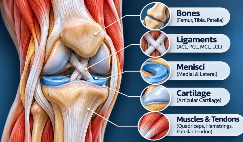
Three bones form the main knee joint: the femur (thighbone), tibia (shinbone), and patella (kneecap). The nearby fibula provides attachment points for supporting structures. Together, these bones and surrounding soft tissues help the knee bear weight and remain stable during walking, running, standing, and squatting. Their arrangement is described in detail in the anatomical overview of the knee.
Knee Anatomy Overview: Main Structures and Functions
The following table summarizes the major components of human knee anatomy, including their locations and functions.
| Structure | Location | Primary Function |
| Femur | Above the knee | Transfers weight through the joint |
| Tibia | Below the knee | Supports weight and forms the lower joint surface |
| Patella | Front of the knee | Protects the joint and improves quadriceps leverage |
| Fibula | Outer lower leg | Provides attachment points for stabilizing structures |
| ACL | Inside the knee | Limits forward movement of the tibia |
| PCL | Inside the knee | Limits backward movement of the tibia |
| MCL | Inner knee | Resists excessive inward collapse |
| LCL | Outer knee | Resists excessive outward bowing |
| Menisci | Between femur and tibia | Distribute loads and improve stability |
| Articular cartilage | Joint surfaces | Reduces friction during movement |
| Quadriceps | Front of thigh | Straightens the knee |
| Hamstrings | Back of thigh | Bend and stabilize the knee |
| Quadriceps tendon | Above kneecap | Connects quadriceps muscles to patella |
| Patellar tendon | Below kneecap | Connects patella to tibia |
Bones and Joints of the Knee
The knee has two main articulations:
- Tibiofemoral joint: Where the rounded medial and lateral femoral condyles meet the tibial plateau.
- Patellofemoral joint: Where the patella glides along a groove at the front of the femur.
The tibial plateau forms the upper surface of the shinbone. Its central intercondylar region provides attachment sites for important ligaments and menisci.
The patella is part of the quadriceps tendon mechanism, helping the quadriceps straighten the knee more efficiently. On the outer side of the joint, the fibular head provides an attachment point for structures such as the lateral collateral ligament.
Although the fibula sits close to the knee, it does not directly form either of the two main knee articulations.
Knee Ligaments: ACL, PCL, MCL, and LCL
Four principal ligaments stabilize the knee by limiting excessive movement. Each controls particular forces that could otherwise make the joint unstable.
Anterior cruciate ligament (ACL): Located centrally inside the knee, the ACL prevents excessive forward movement of the tibia relative to the femur. It also contributes to rotational stability.
Posterior cruciate ligament (PCL): Crossing behind the ACL, the PCL limits excessive backward movement of the tibia.
Medial collateral ligament (MCL): Running along the inner knee, the MCL resists valgus stress, which tends to push the knee inward.
Lateral collateral ligament (LCL): Located on the outer knee, the LCL extends from the femur to the fibular head. It resists varus stress, which tends to push the knee outward.
These four ligaments provide much of the knee’s stability, but they are not the only supporting ligaments. The joint capsule and additional posterolateral structures also help control movement. The main stabilizing roles of the ACL, PCL, MCL, and LCL are explained in the clinical overview of knee ligament injuries.
Menisci and Articular Cartilage
Between the femur and tibia are two fibrocartilaginous structures called menisci:
- Medial meniscus: Located on the inner side of the knee, with a relatively broad C-shaped appearance.
- Lateral meniscus: Located on the outer side, with a more circular shape and greater mobility.
Both menisci spread weight across the joint, absorb some of the forces passing through it, and improve stability. They also help the rounded femoral condyles fit against the relatively flat tibial surface.
Articular cartilage serves a different purpose. It forms a smooth covering over the joint surfaces of the femur, tibia, and patella, allowing the bones to glide against one another with minimal friction. These differences are important to understanding the structure and function of knee cartilage and menisci.
| Tissue | Main Role |
| Ligaments | Connect bones and stabilize joints |
| Tendons | Transmit muscle force to bones |
| Articular cartilage | Provides smooth, low-friction joint surfaces |
| Menisci | Distribute loads and improve joint stability |
Knee Muscles and Tendons
The muscles surrounding the knee do more than produce movement. They also help control the joint as body weight shifts during walking, running, and other activities.
Quadriceps and Hamstrings
The quadriceps femoris consists of four muscles: rectus femoris, vastus medialis, vastus lateralis, and vastus intermedius. Working together, they straighten the knee and help control movement when the leg bears weight.
The hamstrings include the biceps femoris, semitendinosus, and semimembranosus. These muscles bend the knee and help control rotation, particularly when the knee is flexed.
Several smaller muscles also contribute. The gastrocnemius assists with knee flexion, while the popliteus helps initiate bending from a fully extended position. The sartorius and gracilis assist with knee flexion and rotation.
Major Knee Tendons
The quadriceps tendon connects the quadriceps muscles to the patella. Below the kneecap, the patellar tendon, also called the patellar ligament, connects the patella to the tibial tuberosity.
Together, these structures form part of the extensor mechanism that allows the leg to straighten.
The iliotibial band helps support the outer knee. On the inner side, the sartorius, gracilis, and semitendinosus tendons join at the pes anserinus, a shared attachment region on the upper tibia.
Joint Capsule, Synovial Membrane, and Bursae
A strong fibrous capsule surrounds the knee joint. Its inner lining, called the synovial membrane, produces synovial fluid that lubricates the moving surfaces and helps nourish the articular cartilage.
Several small, fluid-filled sacs called bursae reduce friction where tendons, skin, and other tissues move against one another. Important examples include:
- Prepatellar bursa: Located directly in front of the kneecap.
- Suprapatellar bursa: Positioned above the kneecap and normally connected to the joint cavity.
- Infrapatellar bursae: Found around the area below the patella.
- Pes anserine bursa: Located near the inner upper tibia, beneath the pes anserinus tendons.
Fluid inside the knee joint is generally separate from fluid in most surrounding bursae, although certain synovial spaces communicate with the joint cavity.
Nerves and Blood Supply Around the Knee
The area behind the knee is called the popliteal fossa. This region contains major nerves and blood vessels that travel between the thigh and lower leg.
The popliteal artery and vein pass through the fossa. The artery generally lies deeper than the vein and the tibial nerve. Their positions are important because these structures run close together behind the joint.
Branches of the popliteal artery and other nearby vessels form the genicular arterial network, which supplies tissues around the knee.
The tibial nerve passes through the popliteal fossa, while the common fibular nerve travels toward the outer side of the knee and around the fibular neck. These relationships are described in the anatomy of the popliteal region.
The femoral nerve supplies the quadriceps muscles, and its saphenous branch provides sensation to parts of the inner knee and lower leg.
How the Knee Moves and Stays Stable
Knee movement depends on the coordinated action of bones, muscles, ligaments, and cartilage. Although the joint resembles a hinge, its motion is more complex than simple bending and straightening.
During flexion, the femur and tibia move relative to each other through a combination of rolling, gliding, and limited rotation. When the knee extends, the quadriceps provide the main force, while the patella tracks along the groove at the front of the femur.
Near full extension, a small automatic rotation called the screw-home mechanism helps the knee reach a stable position. The direction of this rotation depends on whether the foot is planted or free to move.
Throughout these movements, the menisci distribute loads, ligaments prevent excessive motion, and surrounding muscles provide the active control needed to keep the knee stable.
How Knee Anatomy Relates to Common Injuries
Knowing which structures make up the knee helps explain why different injuries affect movement, stability, and comfort in different ways.
- ACL and PCL tears: Injuries to the central ligaments that control excessive forward and backward movement of the tibia.
- MCL and LCL sprains: Damage to the collateral ligaments that protect the knee against excessive side-to-side stress.
- Meniscus tears: Injuries to the fibrocartilage between the femur and tibia that can interfere with load distribution and joint movement.
- Knee osteoarthritis: Degenerative changes involving articular cartilage and other joint tissues.
- Patellar tendinopathy: A disorder of the patellar tendon, usually associated with discomfort below the kneecap.
- Bursitis: Inflammation of a bursa that may produce swelling and tenderness around the affected area.
- Joint effusion: An accumulation of excess fluid inside the knee joint, often associated with injury or inflammation.
Discomfort can sometimes point toward an affected region, but pain location alone cannot establish a diagnosis. Different structures can produce overlapping symptoms.
Clinicians may use X-rays to examine bones and joint alignment, MRI to assess internal soft tissues and injuries such as a bone bruise, and ultrasound to evaluate certain superficial structures or fluid collections. A knee MRI examination can provide detailed views of ligaments, menisci, cartilage, tendons, and other tissues when further evaluation is needed.
Frequently Asked Questions
Why is the lateral meniscus more mobile than the medial meniscus?
The lateral meniscus has different attachments and is generally less firmly connected to the joint capsule than the medial meniscus. The medial meniscus also has a close attachment to the deep medial collateral ligament, which limits its mobility. These anatomical differences help explain why the two menisci move differently as the knee bends and straightens.
Why can swelling appear above the kneecap after a knee injury?
Swelling above the kneecap may occur when excess joint fluid collects in the suprapatellar recess, a space that communicates with the knee joint cavity. Injury, inflammation, and other joint conditions can cause fluid to accumulate there. However, swelling in this region can have different causes, so its location alone does not identify the underlying problem.