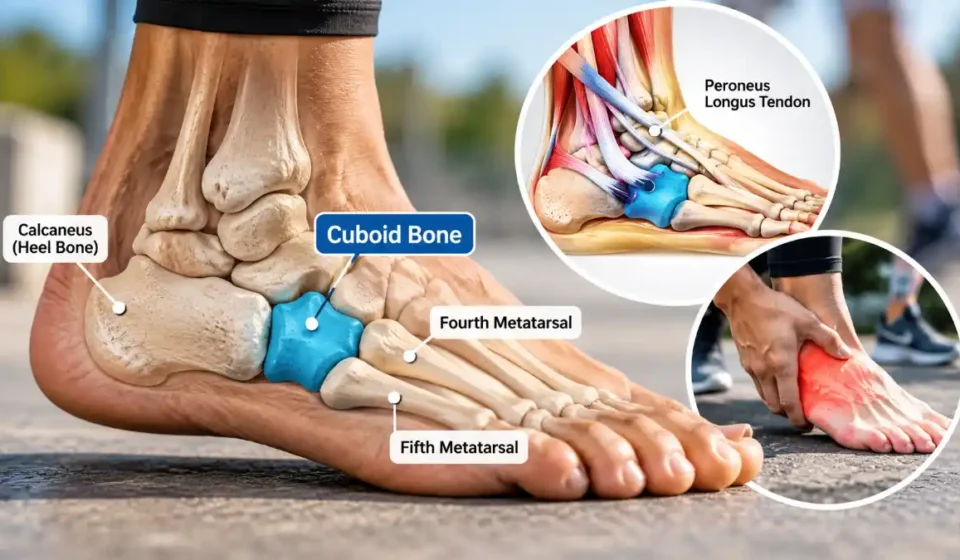

The cuboid bone is a small, roughly cube-shaped tarsal bone on the outer (lateral) side of the midfoot. It sits between the calcaneus (heel bone) and the fourth and fifth metatarsals. This bone helps stabilize the foot, support its arches, and guide the peroneus longus tendon during movement. Despite its small size, the cuboid is important for walking, running, and maintaining balance.
Pain in this area can develop after an ankle sprain, repetitive stress, or a direct injury. Conditions involving the cuboid include cuboid syndrome and fractures, both of which can cause outer midfoot pain, tenderness, swelling, and difficulty walking. Knowing where the bone is located and how it works can help you understand potential injuries and recognize when medical attention is needed.
Where Is the Cuboid Bone Located?
The cuboid bone is located along the outside of the midfoot, just in front of the heel and behind the fourth and fifth metatarsals. These metatarsals extend toward the fourth and fifth toes.
Its main anatomical connections include:
- Behind: The calcaneus, which connects to the cuboid at the calcaneocuboid joint.
- In front: The fourth and fifth metatarsals.
- Toward the inner foot: The lateral cuneiform and, in some people, the navicular bone.
To find its approximate location, feel along the outer edge of your midfoot, just behind the prominent bump at the base of the fifth metatarsal, toward the heel.
However, several bones, tendons, and ligaments lie close together in this area. Feeling pain in a particular spot doesn’t necessarily mean the cuboid itself is injured.
For a closer look at its position and surrounding structures, this cuboid bone anatomy overview explains the bone’s anatomical connections and landmarks.
Cuboid Bone Anatomy and Structure
Cuboid bone anatomy is more complex than its name suggests. Although the bone is roughly cube-shaped, its surfaces are irregular, allowing it to connect with neighboring bones and contribute to foot movement.
Its main anatomical features include:
- Calcaneocuboid joint: Connects the cuboid to the heel bone and helps the midfoot move in a coordinated way.
- Cuboid-metatarsal joints: Connect the cuboid to the fourth and fifth metatarsals, allowing forces to pass through the outer foot.
- Medial articulations: Form connections with the lateral cuneiform and, in some feet, the navicular bone.
- Plantar groove: A channel underneath the cuboid that accommodates the peroneus longus tendon.
The peroneus longus tendon passes beneath the foot through this region. As it changes direction around the cuboid, the bone acts like a pulley, helping the tendon perform its role in foot movement and stability.
Strong surrounding ligaments also help keep the cuboid and neighboring bones stable during everyday activities.
What Does the Cuboid Bone Do?
The main cuboid bone function is to support the outer portion of the foot while allowing controlled movement during activities that involve bearing weight.
Its key functions include:
- Maintaining foot stability: Helps preserve the structure and length of the lateral column, which runs from the heel toward the outer metatarsals.
- Supporting foot arches: Contributes to the lateral longitudinal arch and the mechanics of the transverse arch.
- Transferring forces: Helps distribute mechanical loads through the outer midfoot when standing, walking, or running.
- Supporting movement: Allows coordinated midfoot motion as the foot adjusts to uneven ground and prepares to push off.
- Guiding tendon movement: Redirects the peroneus longus tendon, which contributes to foot stability during activity.
The cuboid doesn’t perform these functions alone. It works with surrounding bones, joints, ligaments, and muscles to keep the foot stable while allowing the flexibility needed for movement.
What Causes Cuboid Bone Pain?
Cuboid bone pain is typically felt along the outer side of the midfoot. It may begin suddenly after an injury or develop gradually from repeated stress.
Possible causes include cuboid syndrome, fractures, ankle sprains, repetitive running or jumping, and injuries involving nearby tendons or ligaments.
Symptoms often include tenderness over the outer midfoot, swelling, discomfort while walking, and pain when pushing off the affected foot. More serious injuries can make it difficult to put weight on that foot.
Not all pain in this area comes from the cuboid. Injuries to the fifth metatarsal, surrounding joints, or peroneal tendons can produce similar symptoms. Following an impact, a bone bruise may also cause pain and tenderness without a complete break in the bone.
Cuboid Syndrome vs. Cuboid Fracture
Cuboid syndrome is thought to involve a disturbance in the normal movement or alignment of the joints around the cuboid. It often develops after an ankle sprain or repeated strain. A cuboid fracture, by contrast, is an actual break in the bone caused by trauma or repetitive stress.
| Feature | Cuboid syndrome | Cuboid fracture |
| Main problem | Altered joint mechanics | Broken bone |
| Common causes | Ankle sprain, repetitive strain | Direct trauma, compression, stress |
| Symptoms | Outer midfoot pain, especially during movement | Local pain, tenderness, possible swelling or bruising |
| Diagnosis | Clinical examination and exclusion of other injuries | Examination and appropriate imaging |
| Treatment | Support, rehabilitation, sometimes professional manipulation | Immobilization, activity restriction, occasionally surgery |
Because their symptoms can overlap, distinguishing these conditions often requires a professional examination.
Cuboid syndrome also has no single definitive diagnostic test. A published review of cuboid syndrome explains that its exact mechanism remains uncertain and that diagnosis generally depends on clinical findings and ruling out other causes of lateral foot pain.
How Are Cuboid Bone Injuries Diagnosed?
Healthcare professionals begin by asking about your symptoms, recent injuries, physical activities, and where the pain occurs.
During a physical examination, they may check for swelling, localized tenderness, restricted movement, and difficulty bearing weight.
Imaging may be recommended depending on the suspected injury:
- X-rays: Often the first imaging test when a foot fracture is suspected.
- CT scans: Can provide a more detailed view of complex fractures, bone displacement, or joint involvement.
- MRI: May help detect stress fractures or injuries that aren’t visible on initial X-rays.
A normal X-ray doesn’t always rule out a cuboid fracture, particularly if the injury is subtle or caused by repetitive stress.
The American College of Radiology’s foot trauma imaging guidelines explain when additional imaging, such as CT or MRI, may be appropriate after an initial examination and X-rays.
Cuboid Bone Injury Treatment and Recovery
Treatment depends on the underlying problem, whether it’s a fracture, a disturbance in joint mechanics, or an injury to the surrounding soft tissues.
For less severe injuries, a healthcare professional may recommend temporarily reducing painful activities, wearing supportive footwear, immobilizing the foot, or starting physical therapy.
Cuboid syndrome may improve with rehabilitation and, in selected cases, manipulation performed by a qualified clinician. However, research supporting specific manipulation techniques remains limited. A suspected fracture should be ruled out before these techniques are considered.
Stable cuboid fractures may require a walking boot or cast, along with restrictions on how much weight you can put on the injured foot. Displaced or complex fractures sometimes need surgery to restore proper joint alignment and preserve the length of the foot’s lateral column.
A medical review of cuboid fracture diagnosis and management discusses why treatment decisions depend on the fracture’s location, displacement, and effect on foot stability.
Recovery time varies with the type and severity of the injury. Some fractures require several weeks of immobilization followed by a gradual return to activity. More complicated injuries may take months before unrestricted movement or sports participation is appropriate.
Recovery from cuboid syndrome also varies, especially when ankle sprains or other injuries are involved.
Avoid trying to manipulate the cuboid yourself or continuing strenuous activities when they cause significant pain.
When Should You Seek Medical Care?
If pain along the outside of your midfoot persists, develops after an injury, or makes walking uncomfortable, arrange an evaluation with a healthcare professional.
Seek urgent medical attention if you experience:
- An inability to bear weight after an injury.
- Severe swelling, worsening pain, or a visible foot deformity.
- Numbness, an unusually cold or pale foot, or other signs of poor circulation.
- An open wound associated with a suspected bone injury.
You can also take steps to reduce your risk of cuboid-related injuries. Wear supportive footwear suited to your activities, increase training intensity gradually, and allow enough recovery time between demanding workouts.
Addressing ankle instability and avoiding excessive repetitive loading may also help protect the outer midfoot.
Frequently Asked Questions
Can you walk with a fractured cuboid bone?
Yes, some people can still walk with a fractured cuboid bone, particularly if the fracture is stable or only slightly displaced. However, being able to walk doesn’t mean the bone is uninjured.
Putting weight on an undiagnosed fracture may make the injury worse or interfere with healing. If you suspect a cuboid fracture, limit weight-bearing and seek medical evaluation rather than trying to walk through the pain.