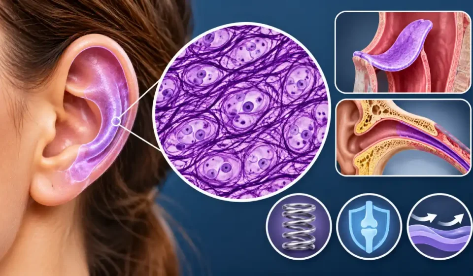

Elastic cartilage is the most flexible of the three main types of cartilage. It provides structural support while allowing tissues to bend and return to their original shape. This specialized connective tissue contains chondrocytes (cartilage cells) housed in small spaces called lacunae, type II collagen, a proteoglycan-rich extracellular matrix, and abundant branching elastic fibers. Together, these components give elastic cartilage its characteristic flexibility while helping it maintain its shape.
Elastic cartilage is found mainly in the external ear, epiglottis, auditory (Eustachian) tube, and certain small laryngeal cartilages. It has no blood vessels of its own, so its cells receive nutrients by diffusion from surrounding tissues. These features distinguish it from other supportive tissues and help explain its limited ability to repair itself.
Where Is Elastic Cartilage Found?
Elastic cartilage supports structures that must retain their shape without becoming rigid. Its main locations include:
- External ear (auricle or pinna): Forms the flexible framework of most of the visible ear, allowing it to bend and return to its usual shape. The soft earlobe contains no cartilage.
- External auditory canal: Supports the cartilaginous outer portion of the ear canal, providing flexibility while helping maintain the canal’s shape.
- Epiglottis: Forms the flexible core of the epiglottis, which moves during swallowing as part of the coordinated process that prevents food and liquid from entering the airway.
- Auditory (Eustachian) tube: Supports the cartilaginous portion of the tube, which opens during activities such as swallowing and yawning to help ventilate the middle ear and equalize pressure.
- Larynx: Forms small structures, including the corniculate and cuneiform cartilages, that provide flexible support within the laryngeal framework.
The National Library of Medicine’s anatomical classification of elastic cartilage identifies the external ear, Eustachian tube, epiglottis, and larynx as its principal locations.
Not every part of these structures contains elastic cartilage. The inner portion of the external auditory canal is bony, for example, while several larger laryngeal cartilages consist primarily of hyaline cartilage.
Elastic Cartilage Structure and Function
Elastic cartilage gets its combination of flexibility and structural support from the organization of its cells, fibers, and extracellular matrix.
Its main components include:
- Chondrocytes: Mature cartilage cells responsible for maintaining the surrounding extracellular matrix.
- Lacunae: Small spaces within the matrix that house chondrocytes, which may occur individually or in small groups.
- Elastic fibers: Branching, interconnected fibers rich in elastin that allow the cartilage to bend and recoil without permanently changing shape under normal conditions.
- Type II collagen: Forms a supportive framework that helps the cartilage withstand mechanical stress.
- Proteoglycan-rich matrix: Holds water and contributes to the tissue’s firmness and resistance to compression.
The elastic fibers are especially important. When you bend the outer ear, their interconnected network helps the cartilage spring back into shape. Collagen and the hydrated matrix provide additional support, helping the tissue resist deformation.
As explained in the University of Leeds’ overview of cartilage structure, the composition of cartilage’s extracellular matrix determines much of its mechanical behavior.
Elastic cartilage has two closely related functions: preserving anatomical shape and allowing flexible movement. Both are important in the external ear, while the epiglottis depends on flexibility to move during swallowing without losing its structural support.
Elastic Cartilage Histology: How to Identify It
Under a microscope, elastic cartilage looks similar to hyaline cartilage in its cellular arrangement. The distinguishing feature is its dense network of branching elastic fibers.
Its main microscopic characteristics include:
- Chondrocytes within lacunae: Round or oval cells sit within spaces in the matrix. They may appear individually or occasionally in small clusters called isogenous groups.
- Branching elastic fibers: Fine, interconnected fibers extend throughout the matrix between lacunae. Special staining makes this network easier to see.
- Perichondrium: A connective tissue covering surrounds the cartilage, helping support its growth and maintenance.
The perichondrium has two layers. The outer fibrous layer contains dense connective tissue and blood vessels, while the inner chondrogenic layer contains cells that can contribute to cartilage formation.
The Digital Histology examination of elastic cartilage shows these features in tissue from the external ear, including the elastic fiber network, chondrocytes, and perichondrial layers.
How Does Elastic Cartilage Appear With Different Stains?
With routine hematoxylin and eosin (H&E) staining, chondrocytes and lacunae are generally easy to identify. However, elastic fibers may not stand out clearly, making elastic cartilage difficult to distinguish from hyaline cartilage.
Histologists use special stains to reveal the elastic fiber network:
- Orcein: Commonly stains elastic fibers reddish-brown.
- Verhoeff’s stain: Typically makes elastic fibers appear dark purple or black.
- Resorcin-fuchsin: Highlights elastic fibers in dark violet or purple tones, depending on the staining method.
These techniques make the branching fibers more visible and help confirm the tissue’s identity.
The Histology Guide’s elastic cartilage specimens demonstrate how the tissue appears with H&E and Verhoeff staining.
For microscopic identification, look for chondrocytes enclosed in lacunae, an extensive elastic fiber network, and a surrounding perichondrium. The fibers are the most reliable distinguishing feature when an appropriate stain is used.
Elastic Cartilage vs. Hyaline Cartilage vs. Fibrocartilage
All three major cartilage types contain chondrocytes embedded in an extracellular matrix. Their differences lie mainly in their fiber composition, flexibility, and ability to withstand particular mechanical forces.
| Feature | Elastic cartilage | Hyaline cartilage | Fibrocartilage |
| Main fibers | Elastic fibers and type II collagen | Primarily type II collagen | Prominent type I collagen, with variable type II |
| Flexibility | Highly flexible with elastic recoil | Firm with limited flexibility | Tough and adapted to heavy loading |
| Main function | Flexible support and shape retention | Structural support, compression resistance, and smooth joint movement | Resistance to compression and tensile stress |
| Chondrocytes | Single cells or small groups in lacunae | Often arranged in small isogenous groups | Commonly arranged in rows between collagen bundles |
| Perichondrium | Present | Usually present, except at joint surfaces and certain other sites | Absent |
| Typical locations | Outer ear, epiglottis, auditory tube | Nose, trachea, articular surfaces | Intervertebral discs, menisci, pubic symphysis |
The clearest difference is the composition and arrangement of the fibers. Elastic cartilage contains abundant elastin-rich fibers that allow bending and recoil. Hyaline cartilage has a smoother, glassy-looking matrix dominated by type II collagen, while fibrocartilage contains thick collagen bundles that help it withstand substantial tensile and compressive forces.
For example, the outer ear contains elastic cartilage because it needs to remain flexible. By contrast, the supporting cartilage of the nose and trachea is predominantly hyaline.
The University of Leeds’ comparison of the three cartilage types explains how their different structures suit their functions.
No single cartilage type is universally stronger or better than the others. Each is adapted to the physical demands of its anatomical location.
How Does Elastic Cartilage Receive Nutrients and Heal?
Elastic cartilage is avascular, meaning it has no blood vessels within the cartilage itself. Instead, oxygen and nutrients diffuse through the extracellular matrix from blood vessels in the surrounding perichondrium.
The perichondrium also supports cartilage growth and contains cells involved in limited tissue repair. However, mature cartilage has a relatively poor capacity for regeneration. Its lack of a direct blood supply and the dense matrix surrounding its cells make recovery from significant damage difficult.
Injuries to the external ear may therefore heal slowly or incompletely, particularly when the perichondrium is damaged, or the cartilage’s nutrient supply is disrupted. This limited healing capacity also helps explain why ear cartilage piercings generally take longer to heal than piercings through the soft earlobe. Inflammation of the perichondrium can further threaten the health of the underlying cartilage.
Although the perichondrium contributes to maintenance and some repair, it cannot guarantee complete cartilage regeneration after a serious injury.